. 2019 Nov 15:202:116086.
doi: 10.1016/j.neuroimage.2019.116086. Epub 2019 Aug 8.

Negative life experiences contribute to racial differences in the neural response to threat

Affiliations

Negative life experiences contribute to racial differences in the neural response to threat

Nathaniel G Harnett et al. Neuroimage. .

Abstract

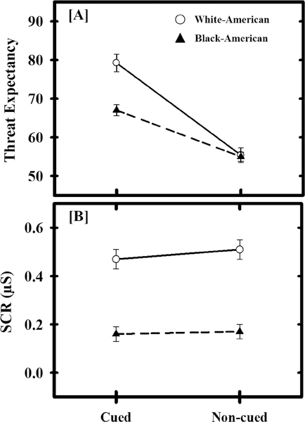
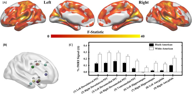
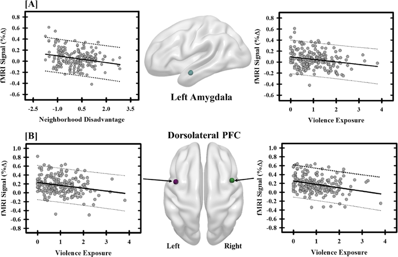
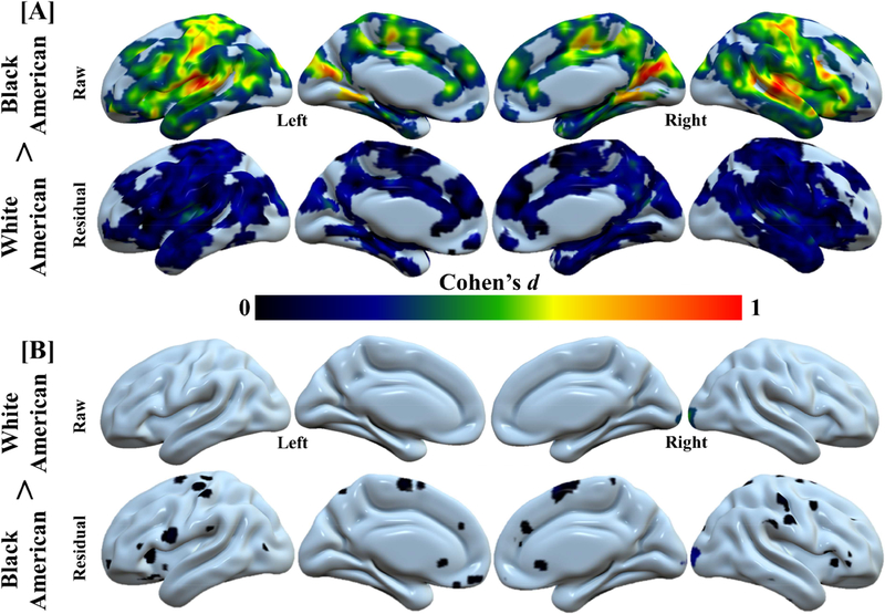
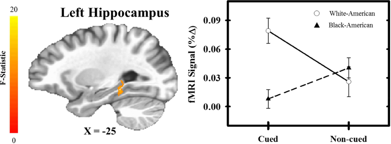
Threat-related emotional function is supported by a neural circuit that includes the prefrontal cortex (PFC), hippocampus, and amygdala. The function of this neural circuit is altered by negative life experiences, which can potentially affect threat-related emotional processes. Notably, Black-American individuals disproportionately endure negative life experiences compared to White-American individuals. However, the relationships among negative life experiences, race, and the neural substrates that support threat-related emotional function remains unclear. Therefore, the current study investigated whether the brain function that supports threat-related emotional processes varies with racial differences in negative life experiences. In the present study, adolescent violence exposure, family income, and neighborhood disadvantage were measured prospectively (i.e., at 11-19 years of age) for Black-American and White-American volunteers. Participants then, as young adults (i.e., 18-23 years of age), completed a Pavlovian fear conditioning task during functional magnetic resonance imaging (fMRI). Cued and non-cued threats were presented during the conditioning task and behavioral (threat expectancy) and psychophysiological responses (skin conductance response; SCR) were recorded simultaneously with fMRI. Racial differences were observed in neural (fMRI activity), behavioral (threat expectancy), and psychophysiological (SCR) responses to threat. These threat-elicited responses also varied with negative life experiences (violence exposure, family income, and neighborhood disadvantage). Notably, racial differences in brain activity to threat were smaller after accounting for negative life experiences. The present findings suggest that racial differences in the neural and behavioral response to threat are due, in part, to exposure to negative life experiences and may provide new insight into the mechanisms underlying racial disparities in mental health.

Keywords: Brain imaging; Fear; Health disparities; Race differences; Social neuroscience; Stress.


Figures

Figure 1.
Figure 1.
Racial differences in behavioral and psychophysiological responses. White-American (White Circles) participants had greater threat expectancy to cued, but not non-cued, threat than Black-American (Black Triangles) participants (a). Further, White-American participants produced larger threat-elicited skin conductance responses (SCR; b) than Black-American participants. Circles (White-Americans) and triangles (Black-Americans) represent mean threat expectancy and SCR for each trial-type and group. Error bars reflect standard error of the mean. SCR data were square-root transformed.
Figure 2.
Figure 2.
Racial differences in the neural response to threat. A significant main effect of race (White-American versus Black-American) was observed across the brain (a). Follow-up analyses were completed on significant peaks of activation in regions important for threat-related emotional function (b). Within these regions, White-American participants (white bars) exhibited a greater fMRI signal response (% change) to threat than Black-American participants (black bars) (c). Bars reflect the mean fMRI signal response for each group and error bars reflect the standard error of the mean. Numbers next to region labels in (c) correspond to numbered regions in (b).
Figure 3.
Figure 3.
Negative life experiences influence threat-elicited brain activity. The fMRI signal response (% change) within the amygdala showed negative relationships with neighborhood disadvantage and violence exposure (a). Specifically, participants with more neighborhood disadvantage and violence exposure showed diminished amygdala activity. Negative relationships were also observed between violence exposure and the fMRI signal response to threat within the dorsolateral prefrontal cortex (PFC) (b). Graphs depict the mean fMRI signal response (% change) and negative life experience (i.e., neighborhood disadvantage and violence exposure) for each participant (grey dots). The solid black line reflects the line of best fit, and the dashed lines represent the prediction bands.
Figure 4.
Figure 4.
Race-related differences in brain activity are accounted for by negative life experience. Negative life experience diminishes the effect of race (i.e., White-American and Black-American) on the neural response to threat. Cohen’s d values from “raw” (i.e., the % fMRI signal responses obtained from the first-level analyses) and “residual” (i.e., the residuals of the model that assessed the relationship between negative life experiences and % fMRI signal responses) neural activity for White-American > Black-American (a) and Black-American > White-American (b) analyses. Large effects of race were observed in the threat-elicited fMRI signal response (i.e., White-American > Black-American: raw). However, these racial differences were reduced (i.e., White-American > Black-American: residual) after accounting for negative life experiences (i.e., violence exposure, family income, and neighborhood disadvantage). A comparison of the confidence intervals for the raw and residual effect sizes is presented in Figure S1.
Figure 5.
Figure 5.
Interaction between race and threat type. A significant interaction effect was observed within the hippocampus such that White-American (White Circles) participants exhibited a greater fMRI signal response (% change) to cued compared to non-cued threat. In contrast, Black-American (Black Triangles) participants exhibited greater fMRI signal responses to non-cued compared to cued threat. Circles (White-Americans) and triangles (Black-Americans) represent mean fMRI signal response for each trial-type and group. Error bars reflect standard error of the mean.

References

    1. Aisenberg E, & Herrenkohl T (2008). Community violence in context: Risk and resilience in children and families. Journal of interpersonal violence, 23(3), 296–315. - PubMed
    1. Alvarez K, Fillbrunn M, Green JG, Jackson JS, Kessler RC, McLaughlin KA,… Alegria M (2018). Race/ethnicity, nativity, and lifetime risk of mental disorders in US adults. Social psychiatry and psychiatric epidemiology, 1–13. - PMC - PubMed
    1. Anderson ER, & Mayes LC (2010). Race/ethnicity and internalizing disorders in youth: a review. Clin Psychol Rev, 30(3), 338–348. doi: 10.1016/j.cpr.2009.12.008 - DOI - PubMed
    1. Atlas LY, Lindquist MA, Bolger N, & Wager TD (2014). Brain mediators of the effects of noxious heat on pain. Pain, 155(8), 1632–1648. doi: 10.1016/j.pain.2014.05.015 - DOI - PMC - PubMed
    1. Baxter R (1966). Diminution and recovery of the UCR in delayed and trace classical GSR conditioning. J Exp Psychol, 71(3), 447–451. - PubMed

Publication types