. 2019;180(2):91-102.
doi: 10.1159/000501637. Epub 2019 Aug 9.

AK002, a Humanized Sialic Acid-Binding Immunoglobulin-Like Lectin-8 Antibody that Induces Antibody-Dependent Cell-Mediated Cytotoxicity against Human Eosinophils and Inhibits Mast Cell-Mediated Anaphylaxis in Mice

Affiliations

AK002, a Humanized Sialic Acid-Binding Immunoglobulin-Like Lectin-8 Antibody that Induces Antibody-Dependent Cell-Mediated Cytotoxicity against Human Eosinophils and Inhibits Mast Cell-Mediated Anaphylaxis in Mice

Bradford A Youngblood et al. Int Arch Allergy Immunol. 2019.

Abstract

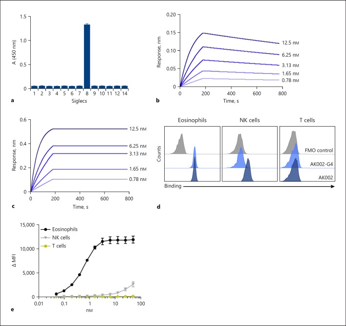
Introduction: Pathologic accumulation and activation of mast cells and eosinophils are implicated in allergic and inflammatory diseases. Sialic acid-binding immunoglobulin-like lectin (Siglec)-8 is an inhibitory receptor selectively expressed on mast cells, eosinophils and, at a lower extent, basophils. When engaged with an antibody, Siglec-8 can induce apoptosis of activated eosinophils and inhibit mast cell activation. AK002 is a humanized, non-fucosylated IgG1 anti-Siglec-8 antibody undergoing clinical investigation for treatment of allergic, inflammatory, and proliferative diseases. Here we examine the human tissue selectivity of AK002 and evaluate the in vitro, ex vivo, and in vivo activity of AK002 on eosinophils and mast cells.

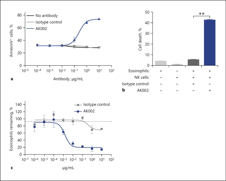
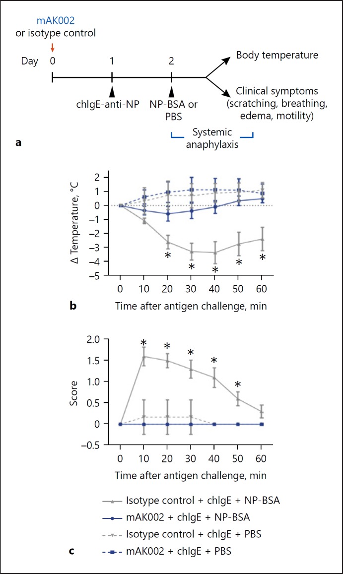
Methods: The affinity of AK002 for Siglec-8 and CD16 was determined by biolayer interferometry. Ex vivo activity of AK002 on human eosinophils from blood and dissociated human tissue was tested in apoptosis and antibody-dependent cell-mediated cytotoxicity (ADCC) assays. The in vivo activity of a murine precursor of AK002 (mAK002) was tested in a passive systemic anaphylaxis (PSA) humanized mouse model.

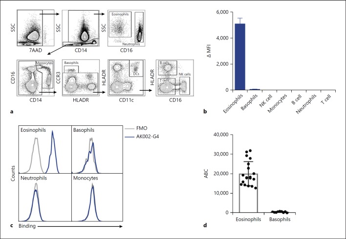
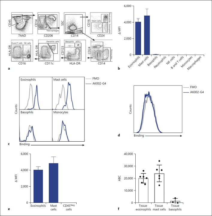
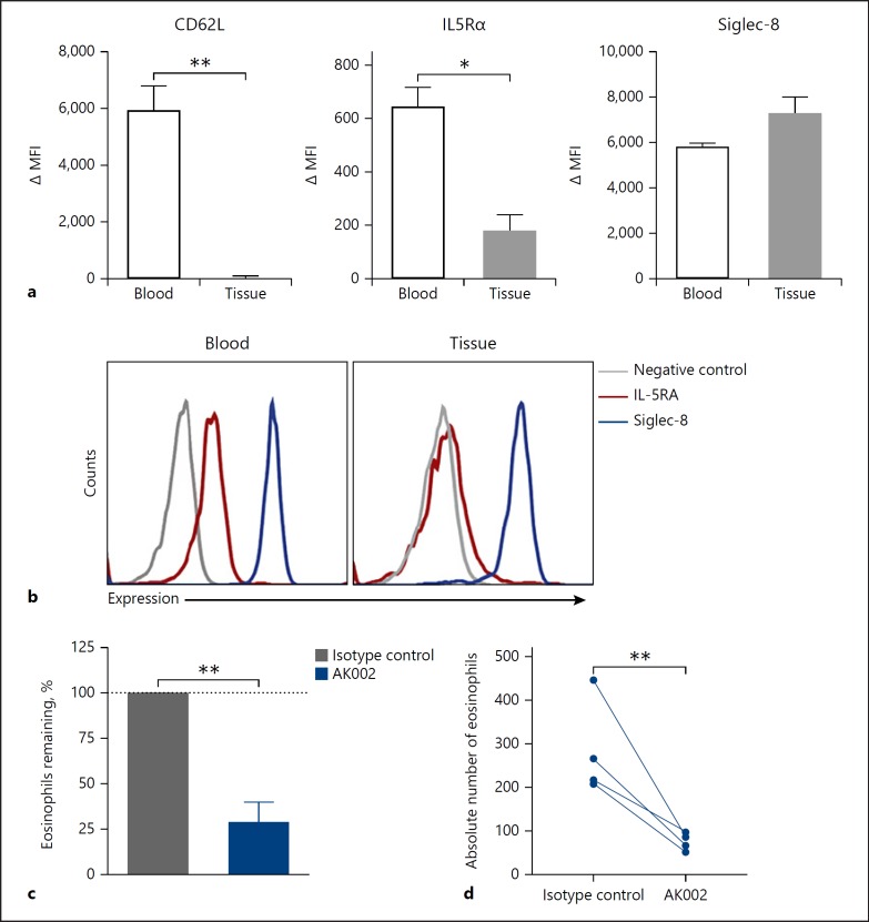
Results: AK002 bound selectively to mast cells, eosinophils and, at a lower level, to basophils in human blood and tissue and not to other cell types examined. AK002 induced apoptosis of interleukin-5-activated blood eosinophils and demonstrated potent ADCC activity against blood eosinophils in the presence of natural killer cells. AK002 also significantly reduced eosinophils in dissociated human lung tissue. Furthermore, mAK002 prevented PSA in humanized mice through mast cell inhibition.

Conclusion: AK002 selectively evokes potent apoptotic and ADCC activity against eosinophils and prevents systemic anaphylaxis through mast cell inhibition.

Keywords: AK002; Anaphylaxis; Antibody-dependent cell-mediated cytotoxicity; Eosinophils; Mast cells; Sialic acid-binding immunoglobulin-like lectin-8.


Conflict of interest statement

Drs. Bradford A. Youngblood, Rustom Falahati, Christopher Bebbington, and Nenad Tomasevic, and Ms. Emily C. Brock and Jessica Bright and Mr. John Leung and Jason Williams are or were employees and have ownership interest in Allakos Inc., outside the submitted work; Drs. Leonard D. Shultz, Dale L. Greiner, and Michael A. Brehm report personal fees from Allakos, during the conduct of the study. Dr. Leonard D. Shultz also reports personal fees from Allakos, outside the submitted work; Drs. Dale L. Greiner and Michael A. Brehm also received grant support and were consultants for The Jackson Laboratory. Dr. Paul J. Bryce has no conflicts of interest to disclose.

Figures

Fig. 1
Fig. 1
AK002 is a non-fucosylated, humanized antibody that is specific for Siglec-8 and interacts with CD16a on NK cells. a AK002 specificity and cross-reactivity was examined using a recombinant human Siglec cross-reactivity ELISA. Recombinant Siglecs were coated on the plate overnight at 0.2 µg/mL and AK002 was added at 2 µg/mL for 2 h. The binding kinetics of (b) monovalent AK002 Fabs or (c) full-length IgG AK002 (two-fold dilutions from 12.5 to 0.8 nM) to the Siglec-8 ECD antigen was measured using biolayer interferometry. The following kinetic parameters were determined for the Fab and full-length IgG, respectively: kon = 1.04 × 10⁶ 1/Ms, koff = 4.82 × 10⁻⁴ 1/s, Kd = 464 pM and kon = 1.86 × 10⁶ 1/Ms, since no significant dissociation was observed for the measured time of 600 s Kd was assigned to <1 pM. d Binding of 50 nM AK002-G4 (light blue) and AK002 (dark blue) to eosinophils, NK cells, and T cells gated as shown in (c) in human peripheral blood by flow cytometry. e Titration of AK002 binding in human peripheral blood to eosinophils, NK cells, and T cells gated as shown in panel C by flow cytometry. Siglec, sialic acid-binding immunoglobulin-like lectin; NK, natural killer; MFI, median fluorescence intensity; FMO, fluorescence minus one.
Fig. 2
Fig. 2
AK002-G4 selectively binds to eosinophils and basophils in human peripheral blood. a Flow cytometry gating strategy used to identify immune cell subtypes among human PBL. b AK002-G4 binding plotted as ΔMFI on immune cells identified using gating strategy in a (mean ± SD of 4 donors). c Representative histograms for eosinophils, basophils, neutrophils, and monocytes stained with an FMO control (gray) or AK002-G4 (blue). d Siglec-8 ABC of peripheral blood eosinophils (median 18,029 ± 6,133) and basophils (median 552 ± 292) determined by quantitative cytometry (individual donors are plotted ± SD). ΔMFI was determined by subtracting the MFI for a FMO control sample from the MFI for cells stained with the conjugated antibody. ΔMFI values that were negative after subtracting the FMO were given a value of zero. ABC, antibody binding capacity; MFI, median fluorescence intensity; FMO, fluorescence minus one; NK, natural killer.
Fig. 3
Fig. 3
AK002-G4 selectively binds to eosinophils and mast cells from human tissue. a Flow cytometry gating strategy used to identify immune cell subtypes from human lung tissue. b AK002-G4 binding plotted as ΔMFI on immune cells identified using gating strategy in (a; Mean ± SD of 4 donors). c Representative histograms for eosinophils, mast cells, basophils, and monocytes stained with an FMO control (gray) or AK002-G4 (blue). d Representative histogram for viable CD45-negative cells in human lung tissue gated as shown above stained with an FMO control (gray) or AK002-G4 (blue). e AK002-G4 binding plotted as ΔMFI on eosinophils, mast cells and viable, CD45 negative cells in human lung tissue (mean ± SD of 4 donors). f Siglec-8 ABC of lung tissue eosinophils (median 21,529 ± 7,397), lung tissue mast cells (median 22,246 ± 7,929), and lung tissue basophils (median 621 ± 2,085) determined by quantitative cytometry (individual donors are plotted ± SD). ΔMFI was determined by subtracting the MFI for an FMO control sample from the MFI for cells stained with the conjugated antibody. ΔMFI values that were negative after subtracting the FMO were given a value of zero. MFI, median fluorescence intensity; FMO, fluorescence minus one; NK, natural killer; ABC, antibody binding capacity.
Fig. 4
Fig. 4
AK002 has potent apoptosis-inducing activity and ADCC activity on human eosinophils. a IL-5 activated purified human eosinophils incubated overnight with AK002 (blue) or isotype control antibody (gray) followed by apoptosis detection using Annexin V-PE and 7AAD (mean ± SD of 4 biological replicates). b Purified peripheral blood eosinophils cultured with purified NK cells at a 9:1 effector:target cell ratio in the presence of 10 μg/mL isotype control or AK002 for 4 h. Cell death determined by lactate dehydrogenase release in cell supernatants (mean ± SD of 3 biological replicates). c PBL from healthy donor incubated overnight with AK002 (blue) or isotype control antibody (gray) and percentage of eosinophils remaining determined (mean ± SD of 3 technical replicates; representative data of 3 donors). In (c), eosinophils were counted by flow cytometry and the percent eosinophils remaining was calculated by normalizing to the percent of CD45+ eosinophils in the isotype control-treated wells to 100%. * p < 0.05; ** p < 0.01. NK, natural killer.
Fig. 5
Fig. 5
AK002 reduces human tissue eosinophils ex vivo. a Surface expression of CD62L, IL-5Rα and Siglec-8 plotted as ΔMFI on blood (black) and lung tissue (gray) eosinophils (mean ± SD of 4 donors). b Representative histograms of surface expression of IL-5Rα (red), Siglec-8 (blue) or fluorescence minus one negative control (gray) on blood and lung tissue eosinophils. c, d Dissociated human tissue was incubated overnight with 1 μg/mL isotype control (gray) or AK002 (blue). Eosinophils were counted by flow cytometry and plotted as the (c) percent eosinophils remaining or as (d) absolute eosinophil counts. The percent eosinophils remaining was calculated by normalizing to the percent of CD45+ eosinophils in the isotype control-treated wells to 100% (mean ± SD of 4 donors). * p < 0.05; ** p < 0.01. MFI, median fluorescence intensity; IL5Rα, interleukin-5 receptor α; Siglec, sialic acid-binding immunoglobulin-like lectin.
Fig. 6
Fig. 6
mAK002 inhibits IgE-mediated PSA in humanized mice. a Experimental design of PSA in NSG-SGM3-BLT humanized mice. NSG-SGM3 BLT mice were treated with either 100 μg of isotype control antibody (gray) or mAK002 (blue). The next day, the mice were systemically sensitized with chIgE-anti-NP antibody and systemically challenged 24 h later with BSA-NP (solid line) or PBS (dashed line). Systemic anaphylaxis was measured by monitoring the rectal body temperature (b, c) observable clinical symptom scores as described in methods (mean ± SD of 2 experiments; n = 5–10 mice/group). * p < 0.05. BSA, bovine serum albumin; chIgE, chimeric human IgE; NP, nitrophenacetyl; PBS, phosphate-buffered saline.

References

    1. Hudson SA, Herrmann H, Du J, Cox P, Haddad B, Butler B, et al. Developmental, malignancy-related, and cross-species analysis of eosinophil, mast cell, and basophil siglec-8 expression. J Clin Immunol. 2011 Dec;31((6)):1045–53. - PMC - PubMed
    1. Kikly KK, Bochner BS, Freeman SD, Tan KB, Gallagher KT, D'alessio KJ, et al. Identification of SAF-2, a novel siglec expressed on eosinophils, mast cells, and basophils. J Allergy Clin Immunol. 2000 Jun;105((6 Pt 1)):1093–100. - PubMed
    1. Floyd H, Ni J, Cornish AL, Zeng Z, Liu D, Carter KC, et al. Siglec-8. A novel eosinophil-specific member of the immunoglobulin superfamily. J Biol Chem. 2000 Jan;275((2)):861–6. - PubMed
    1. Yokoi H, Choi OH, Hubbard W, Lee HS, Canning BJ, Lee HH, et al. Inhibition of FcepsilonRI-dependent mediator release and calcium flux from human mast cells by sialic acid-binding immunoglobulin-like lectin 8 engagement. J Allergy Clin Immunol. 2008 Feb;121((2)):499–505.e1. - PubMed
    1. Nutku E, Aizawa H, Hudson SA, Bochner BS. Ligation of Siglec-8: a selective mechanism for induction of human eosinophil apoptosis. Blood. 2003 Jun;101((12)):5014–20. - PubMed

MeSH terms