Review
. 2019 Aug 10;19(9):83.
doi: 10.1007/s11892-019-1194-6.

Alterations in Beta Cell Identity in Type 1 and Type 2 Diabetes

Affiliations
Review

Alterations in Beta Cell Identity in Type 1 and Type 2 Diabetes

Abu Saleh Md Moin et al. Curr Diab Rep. .

Abstract

Purpose of review: To discuss the current understanding of "β cell identity" and factors underlying altered identity of pancreatic β cells in diabetes, especially in humans.

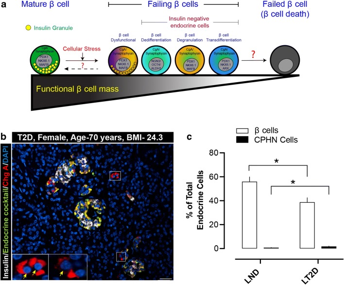
Recent findings: Altered identity of β cells due to dedifferentiation and/or transdifferentiation has been proposed as a mechanism of loss of β cells in diabetes. In dedifferentiation, β cells do not undergo apoptosis; rather, they lose their identity and function. Dedifferentiation is well characterized by the decrease in expression of key β cell markers such as genes encoding major transcription factors, e.g., MafA, NeuroD1, Nkx6.1, and Foxo1, and an increase in atypical or "disallowed" genes for β cells such as lactate dehydrogenase, monocarboxylate transporter MCT1, or progenitor cell genes (Neurog3, Pax4, or Sox9). Moreover, altered identity of mature β cells in diabetes also involves transdifferentiation of β cells into other islet hormone producing cells. For example, overexpression of α cell specific transcription factor Arx or ablation of Pdx1 resulted in an increase of α cell numbers and a decrease in β cell numbers in rodents. The frequency of α-β double-positive cells was also prominent in human subjects with T2D. These altered identities of β cells likely serve as a compensatory response to enhance function/expand cell numbers and may also camouflage/protect cells from ongoing stress. However, it is equally likely that this may be a reflection of new cell formation as a frank regenerative response to ongoing tissue injury. Physiologically, all these responses are complementary. In diabetes, (1) endocrine identity recapitulates the less mature/less-differentiated fetal/neonatal cell type, possibly representing an adaptive mechanism; (2) residual β cells may be altered in their subtype proportions or other molecular features; (3) in humans, "altered identity" is a preferable term to dedifferentiation as their cellular fate (differentiated cells losing identity or progenitors becoming more differentiated) is unclear as yet.

Keywords: Dedifferentiation; Pancreas; Transdifferentiation; Type 1 diabetes; Type 2 diabetes; β Cell.


Conflict of interest statement

The authors declare that they have no conflict of interest.

Figures

Fig. 1
Fig. 1
a A schematic illustrating the possible “altered differentiation” states of human pancreatic β cells. In response to cellular stresses (glucotoxicity, ER stress, immune attack or viral infection), β cells can undergo functional or morphological changes. These alterations might include insulin-positive dysfunctional β cells or insulin-negative β cells that are dedifferentiated (expressing transcription factors of endocrine progenitor cells), degranulated (“empty” β cells no longer harboring a normal complement of insulin granules), or transdifferentiated (transitioning towards a different endocrine cell subtype). At this stage, the β cells may be recognized as “failing β cells,” as they do not contain any releasable insulin. The unanswered questions are whether human β cells from those transition states can revert back towards the mature, fully functional state, or whether they will eventually fail completely and undergo apoptotic death. b Example of chromograninA-positive hormone-negative (CPHN) cells in the pancreas of a lean subject with type 2 diabetes (Age, 70 years, BMI, 24.3). The merged immunofluorescent image is a selected field of 4-μm paraffin section of pancreas stained for insulin (white), endocrine cocktail (glucagon, somatostatin, pancreatic polypeptide and ghrelin) (green), chromograninA (ChgA) (red), and DAPI (blue). The cells staining only for chromograninA and not for any of the known pancreatic hormones (CPHN cells) are therefore stained red (and indicated by yellow arrows). Inset, high-power image of the selected area (marked by white square in the low power image) indicating the CPHN cells. Scale bar, 50 μm. c Percent of β cells and CPHN cells (of total endocrine cells) in lean human subjects with no diabetes (LND) and lean human subjects with type 2 diabetes (LT2D). Data are presented as mean ± SEM, N = 10 (each group). *, p < 0.01

References

    1. Meier JJ, Bhushan A, Butler AE, Rizza RA, Butler PC. Sustained beta cell apoptosis in patients with long-standing type 1 diabetes: indirect evidence for islet regeneration? Diabetologia. 2005;48(11):2221–2228. doi: 10.1007/s00125-005-1949-2. - DOI - PubMed
    1. Butler AE, Janson J, Bonner-Weir S, Ritzel R, Rizza RA, Butler PC. Beta-cell deficit and increased beta-cell apoptosis in humans with type 2 diabetes. Diabetes. 2003;52(1):102–110. doi: 10.2337/diabetes.52.1.102. - DOI - PubMed
    1. Talchai C, Xuan S, Lin HV, Sussel L, Accili D. Pancreatic beta cell dedifferentiation as a mechanism of diabetic beta cell failure. Cell. 2012;150(6):1223–1234. doi: 10.1016/j.cell.2012.07.029. - DOI - PMC - PubMed
    1. Spijker HS, Ravelli RB, Mommaas-Kienhuis AM, van Apeldoorn AA, Engelse MA, Zaldumbide A, et al. Conversion of mature human beta-cells into glucagon-producing alpha-cells. Diabetes. 2013;62(7):2471–2480. doi: 10.2337/db12-1001. - DOI - PMC - PubMed
    1. Pipeleers D, Chintinne M, Denys B, Martens G, Keymeulen B, Gorus F. Restoring a functional beta-cell mass in diabetes. Diabetes Obes Metab. 2008;10(Suppl 4):54–62. doi: 10.1111/j.1463-1326.2008.00941.x. - DOI - PubMed

MeSH terms