. 2019 Oct 1;27(10):1485-1496.e4.
doi: 10.1016/j.str.2019.07.012. Epub 2019 Aug 8.

Flexible Tethering of ASPP Proteins Facilitates PP-1c Catalysis

Affiliations

Flexible Tethering of ASPP Proteins Facilitates PP-1c Catalysis

Yeyun Zhou et al. Structure. .

Abstract

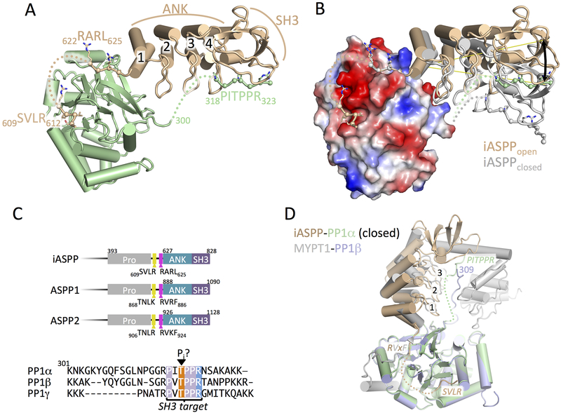
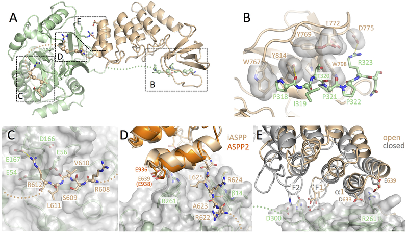
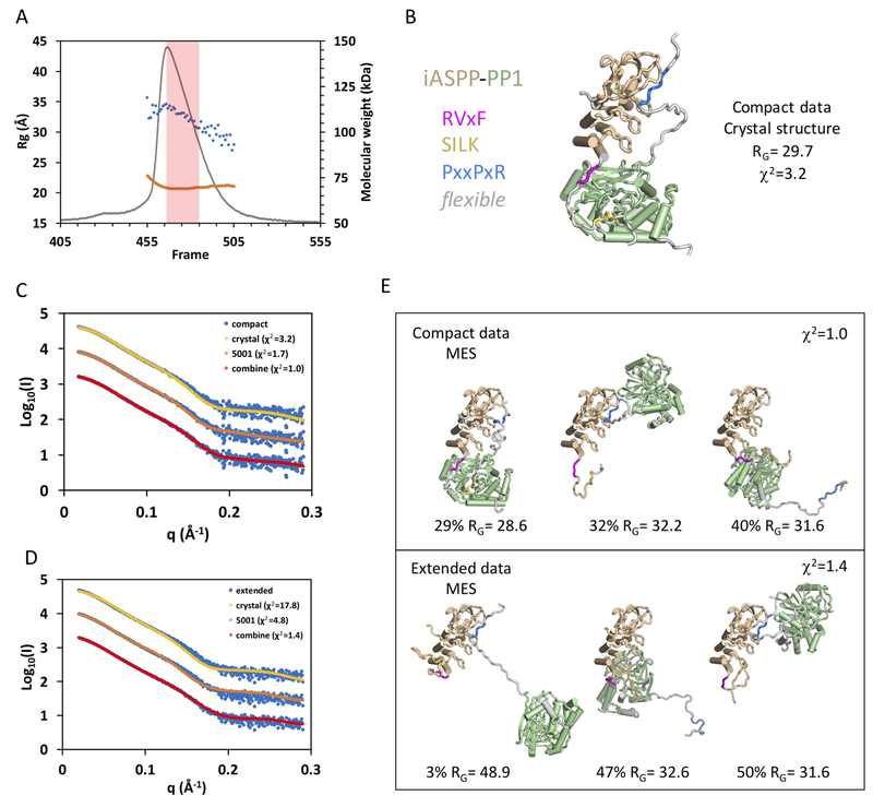
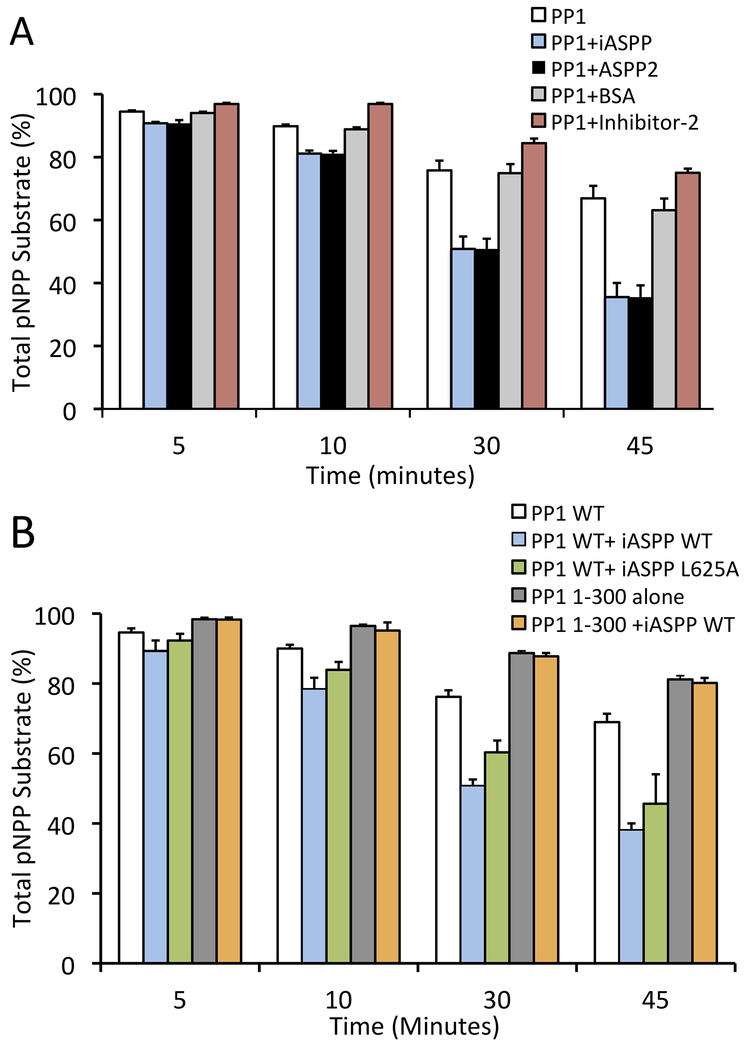
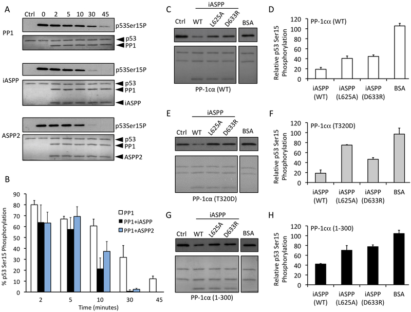
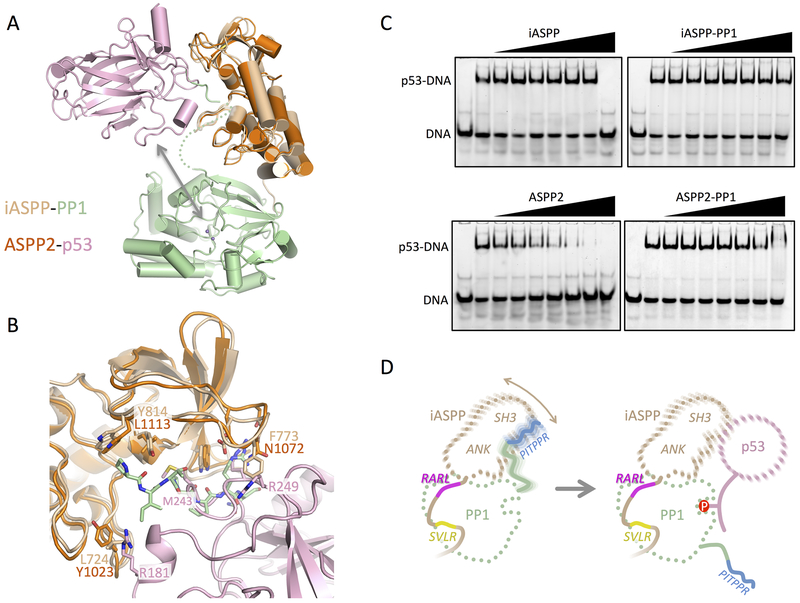
ASPP (apoptosis-stimulating proteins of p53) proteins bind PP-1c (protein phosphatase 1) and regulate p53 impacting cancer cell growth and apoptosis. Here we determine the crystal structure of the oncogenic ASPP protein, iASPP, bound to PP-1c. The structure reveals a 1:1 complex that relies on interactions of the iASPP SILK and RVxF motifs with PP-1c, plus interactions of the PP-1c PxxPxR motif with the iASPP SH3 domain. Small-angle X-ray scattering analyses suggest that the crystal structure undergoes slow interconversion with more extended conformations in solution. We show that iASPP, and the tumor suppressor ASPP2, enhance the catalytic activity of PP-1c against the small-molecule substrate, pNPP as well as p53. The combined results suggest that PxxPxR binding to iASPP SH3 domain is critical for complex formation, and that the modular ASPP-PP-1c interface provides dynamic flexibility that enables functional binding and dephosphorylation of p53 and other diverse protein substrates.

Keywords: ANK repeats; ASPP2; PP-1c; RVxF motif; SH3; Small angle X-ray scattering; X-ray crystallography; dephosphorylation; iASPP; p53.


Conflict of interest statement

DECLARATION OF INTERESTS

The authors declare no competing financial interests.

Figures

Figure 1.
Figure 1.
Crystal structure of iASPP608–828 bound to PP-1cα. (A) Overview of the structure of iASPP608–828 bound to PP-1cα. iASPP is brown, PP-1cα is in green. Peptide segments involved in key protein-protein contacts are illustrated with Cα spheres and the four ANK repeats are numbered. Regions of polypeptide chain not visible in the electron density are indicated by dotted lines. (B) Comparison of the two iASPP608–828-PP-1cα complexes in the asymmetric unit, aligned on PP-1cα, with the second iASPP in the closed conformation colored grey and an electrostatic charge surface displayed for PP-1cα. The ~22° angular difference in the orientation of the two iASPPs relative to PP-1cα is indicated. (C) Top panel, comparison of the primary structures of the C-terminal regions of iASPP, ASPP1 and ASPP2 with key domains and motifs indicated. Bottom panel, sequence alignment of the C-terminal unstructured tails of the three PP-1cα isoforms. (D) Structural comparison of the closed complex of iASPP-PP-1cα with the structure of MYPT1-PP-1β, aligned on PP-1cα with the different chains colored as indicated in the figure. Crystallographic statistics are given in Table 1.
Figure 2.
Figure 2.
Four distinct contact surfaces stabilize the iASPP-PP-1c complex. (A) Overview of the iASPP608–828-PP-1cα structure with the four intermolecular contact surfaces boxed and labeled corresponding to the subsequent panels. (B) Details of the SH3-PxxPxR motif interaction. Key residues from the iASPP SH3 domain that interact with the PxxPxR motif are shown as sticks with transparent surfaces. The PxxPxR peptide is shown in sticks with Cα spheres displayed. (C) Details of the SILK-PP-1c interaction. A transparent surface is shown for PP-1c with key side chains shown as sticks. The SILK peptide is shown in sticks with Cα spheres displayed. (D) Details of the RARL-PP-1c interaction and ANK-PP-1c contact. The RARL peptide is shown in sticks with Cα spheres displayed, while PP-1c is displayed with a transparent surface and key contact side chains as sticks. The ASPP2 structure (PDB ID: 1YCS) is shown aligned on the iASPP structure with conserved acidic residues in the first ANK repeat shown as sticks. (E) Interactions between the iASPP ANK repeats and PP-1c. Shown are the two iASPP-PP-1c complexes aligned on PP-1c. The fingers from the first two ankyrin repeats are labeled F1 and F2 and residues that make intermolecular contacts are shown as sticks. Electron density for each of these contact surfaces is shown in Figure S1.
Figure 3.
Figure 3.
Conformational flexibility of iASPP-PP-1c revealed by SAXS. (A) SEC-SAXS chromatogram with raw scattering trace in grey, the blue dots give the RG values calculated by Guinier approximation for each frame, and the orange trace gives the MW as determined by MALS. (B) MES modeling using the 5011 library (based on the open form of the iASPP-PP-1c crystal structure). The single model is shown with color coding as shown on the left. (C) and (D) Fit of the compact (C) and extended (D) data sets to calculated scattering from the MES models derived from the 5011 library (crystal), the 5001 library, and the combined libraries. (E) Three model ensembles derived from the combined libraries for the compact data set (top panel) and the extended data set (bottom panel). Additional details and a comparable analysis of ASPP2-PP-1c and iASPP(621-828)-PP-1c are given in Figures S2, S3, S4 and S5 and Tables S1 and S2.
Figure 4.
Figure 4.
iASPP and ASPP2 enhance the activity of PP-1c towards pNPP and inhibit PP-1cα towards Phosphorylase a. (A) PP-1cα colorimetric assay using para-nitrophenylphosphate (pNPP) as a substrate for PP-1cα. PP-1cα was incubated alone or in the presence of iASPP608–828, ASPP2905–1128, bovine serum albumin (BSA) or Inhibitor-2. Graph depicts the total amount of pNPP substrate left in each sample at each time point. Error bars indicate s.d of 3 replicates. (B) PP-1c colorimetric pNPP assay comparing the activity of PP-1cα WT or PP-1cα1-300 alone, or in the presence of WT iASPP608–828, or iASPP608–828 L625A mutant. Error bars indicate s.d of 3 replicates.
Figure 5.
Figure 5.
iASPP and ASPP2 enhance the dephosphorylation of p53 Ser-15. (A) Western blot analysis of in vitro dephosphorylation reactions of DNA-PK phosphorylated p532–293 (blotting for phosho-Ser-15) incubated with PP-1cα alone (top panel) or in the presence of iASPP608–828 (middle panel) or ASPP2905–1128 (lower panel) for 0 to 45 min. The lanes labeled “ctrl” contain a sample of p53 where DNA-PK was inhibited with LY294002 for the duration of the experiment. The lanes labeled “0” contain no PP1 or ASPP-PP1. (B) Quantification of the amount of phosphorylated p53 Ser-15 in each sample relative to the total phospho-Ser-15 in control. Error bars indicate s.d. of 4 replicates. (C-H) Western blot analyses of in vitro dephosphorylation reactions of DNA-PK phosphorylated p53 (blotting for phospho-Ser-15 p53) by PP-1cα WT (C and D), PP-1cα T320D (E and F), or PP-1cα1–300 (G and H) in the presence of iASPP WT, iASPP L625A, or iASPP D633R, with results quantitated as in (B). Error bars indicate s.d. of 3 replicates.
Figure 6.
Figure 6.
ASPP-PP-1c-p53 interactions and a model for the targeted dephosphorylation of p53 by ASPP-PP-1c. (A) Interactions between ASPP-PP-1cα complexes and p53. Shown is a structural overlay of the closed iASPP-PP-1cα complex and the ASPP2-p53 DBD complex (PDB ID: 1YCS), aligned on their ASPP components. The arrow indicates the proximity of the p53 DBD to the PP-1c active site catalytic metal ions (purple spheres). (B) Details of the superposition of the iASPP-PP-1c and ASPP2-p53 DBD complex. Shown is the ASPP SH3 domain and C-terminal ANK finger with the p53 DBD and PP-1c PxxPxR peptide shown. Key residues involved in intermolecular contacts are shown as sticks. (C) EMSA analysis probing the ability of ASPP or ASPP- PP-1cα complexes to inhibit p53 DBD binding to a PUMA p53 DNA site. Each lane contains 80 nM PUMA. In each panel, lanes 2–9 contain 800 nM p53 DBD, and a gradient of ASPP protein or ASPP- PP-1cα complex at 0, 0.8, 1.6, 3.2, 6.4, 12.8, 25.6, and 51.2 μM. (D) Model for how a dynamic ASPP-PP-1c complex could bind and target p53 for dephosphorylation. Left panel – the iASPP-PP-1c complex is stabilized by discrete interactions involving the SVLR, RARL and PITPPR peptide motifs, resulting in a flexible complex that allows motion of the iASPP ANK/SH3 domain relative to the PP-1c catalytic domain. Right panel – dynamic release of the PP-1c PITPPR motif allows binding of p53 to the ANK/SH3 domain and dephosphosphorylation.

Comment in

References

    1. Aceytuno RD, Piett CG, Havali-Shahriari Z, Edwards RA, Rey M, Ye R, Javed F, Fang S, Mani R, Weinfeld M, et al. (2017). Structural and functional characterization of the PNKP-XRCC4-LigIV DNA repair complex. Nucleic Acids Res 45, 6238–6251. - PMC - PubMed
    1. Adams PD, Afonine PV, Bunkoczi G, Chen VB, Davis IW, Echols N, Headd JJ, Hung LW, Kapral GJ, Grosse-Kunstleve RW, et al. (2010). PHENIX: a comprehensive Python-based system for macromolecular structure solution. Acta crystallographica Section D, Biological crystallography 66, 213–221. - PMC - PubMed
    1. Agirre X, Roman-Gomez J, Jimenez-Velasco A, Garate L, Montiel-Duarte C, Navarro G, Vazquez I, Zalacain M, Calasanz MJ, Heiniger A, et al. (2006). ASPP1, a common activator of TP53, is inactivated by aberrant methylation of its promoter in acute lymphoblastic leukemia. Oncogene 25, 1862–1870. - PubMed
    1. Ahn J, Byeon IJ, Byeon CH, and Gronenborn AM (2009). Insight into the structural basis of pro- and antiapoptotic p53 modulation by ASPP proteins. The Journal of biological chemistry 284, 13812–13822. - PMC - PubMed
    1. Ao Y, Rohde LH, and Naumovski L (2001). p53-interacting protein 53BP2 inhibits clonogenic survival and sensitizes cells to doxorubicin but not paclitaxel-induced apoptosis. Oncogene 20, 2720–2725. - PubMed

Publication types

MeSH terms