

Non-covalent NRF2 Activation Confers Greater Cellular Protection than Covalent Activation
- PMID: 31402317
- PMCID: PMC6800637
- DOI: 10.1016/j.chembiol.2019.07.011
Non-covalent NRF2 Activation Confers Greater Cellular Protection than Covalent Activation
Abstract
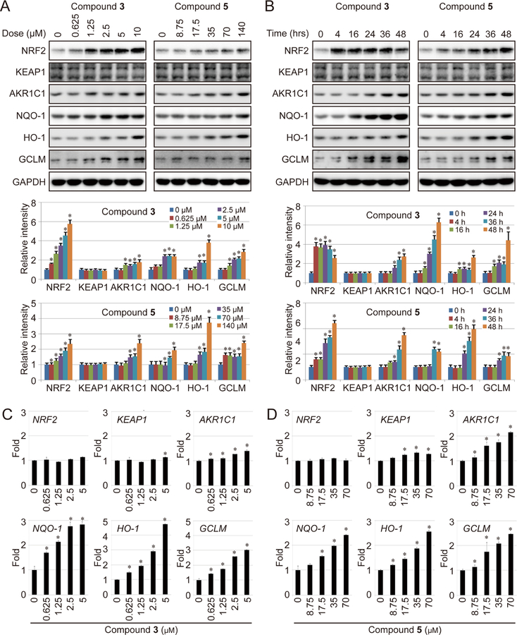
The transcription factor NRF2 confers cellular protection by maintaining cellular redox homeostasis and proteostasis. Basal NRF2 levels are normally low due to KEAP1-mediated ubiquitylation and subsequent proteasomal degradation. KEAP1, a substrate adaptor protein of a KEAP1-CUL3-RBX1 E3 ubiquitin ligase complex, contains a critical cysteine (C151) that is modified by electrophiles or oxidants, resulting in inactivation of the E3 ligase and inhibition of NRF2 degradation. Currently, nearly all NRF2 inducers are electrophilic molecules that possess unwanted off-target effects due to their reactive nature. Here, we report a group of NRF2 inducers, ent-kaurane diterpenoid geopyxins, with and without C151 reactive electrophilic moieties. Among 16 geopyxins, geopyxin F, a non-electrophilic NRF2 activator, showed enhanced cellular protection relative to an electrophilic NRF2 activator, geopyxin C. To our knowledge, this is the first detailed structure-activity relationship study of covalent versus non-covalent NRF2 activators, showing the promise of non-covalent NRF2 activators as potential therapeutic compounds.
Keywords: NRF2; cancer; chemoprevention; drug discovery; geopyxin; natural product; non-covalent; structure activity relationship.
Copyright © 2019 Elsevier Ltd. All rights reserved.
Conflict of interest statement
DECLARATION OF INTERESTS
The authors declare no competing interests.
Figures


Similar articles
-
Review of molecular mechanisms involved in the activation of the Nrf2-ARE signaling pathway by chemopreventive agents.Methods Mol Biol. 2010;647:37-74. doi: 10.1007/978-1-60761-738-9_3. Methods Mol Biol. 2010. PMID: 20694660 Review.
-
The Keap1-Nrf2 system as an in vivo sensor for electrophiles.Nitric Oxide. 2011 Aug 1;25(2):153-60. doi: 10.1016/j.niox.2011.02.007. Epub 2011 Mar 6. Nitric Oxide. 2011. PMID: 21385624
-
Oxidative stress sensor Keap1 functions as an adaptor for Cul3-based E3 ligase to regulate proteasomal degradation of Nrf2.Mol Cell Biol. 2004 Aug;24(16):7130-9. doi: 10.1128/MCB.24.16.7130-7139.2004. Mol Cell Biol. 2004. PMID: 15282312 Free PMC article.
-
Oridonin confers protection against arsenic-induced toxicity through activation of the Nrf2-mediated defensive response.Environ Health Perspect. 2008 Sep;116(9):1154-61. doi: 10.1289/ehp.11464. Environ Health Perspect. 2008. PMID: 18795156 Free PMC article.
-
Molecular mechanisms of the Keap1–Nrf2 pathway in stress response and cancer evolution.Genes Cells. 2011 Feb;16(2):123-40. doi: 10.1111/j.1365-2443.2010.01473.x. Genes Cells. 2011. PMID: 21251164 Review.
Cited by
-
The intricacies of NRF2 regulation in cancer.Semin Cancer Biol. 2021 Nov;76:110-119. doi: 10.1016/j.semcancer.2021.05.016. Epub 2021 May 18. Semin Cancer Biol. 2021. PMID: 34020028 Free PMC article. Review.
-
α-Lipoic Acid Targeting PDK1/NRF2 Axis Contributes to the Apoptosis Effect of Lung Cancer Cells.Oxid Med Cell Longev. 2021 Jun 4;2021:6633419. doi: 10.1155/2021/6633419. eCollection 2021. Oxid Med Cell Longev. 2021. PMID: 34211631 Free PMC article.
-
Targeting Nrf2-Mediated Oxidative Stress Response Signaling Pathways as New Therapeutic Strategy for Pituitary Adenomas.Front Pharmacol. 2021 Mar 24;12:565748. doi: 10.3389/fphar.2021.565748. eCollection 2021. Front Pharmacol. 2021. PMID: 33841137 Free PMC article.
-
The role of natural products in revealing NRF2 function.Nat Prod Rep. 2020 Jun 1;37(6):797-826. doi: 10.1039/c9np00061e. Epub 2020 May 13. Nat Prod Rep. 2020. PMID: 32400766 Free PMC article. Review.
-
Insight into Nrf2: a bibliometric and visual analysis from 2000 to 2022.Front Genet. 2023 Sep 15;14:1266680. doi: 10.3389/fgene.2023.1266680. eCollection 2023. Front Genet. 2023. PMID: 37779908 Free PMC article.
References
-
- Baird L, Lleres D, Swift S, and Dinkova-Kostova AT (2013). Regulatory flexibility in the Nrf2-mediated stress response is conferred by conformational cycling of the Keap1-Nrf2 protein complex. Proceedings of the National Academy of Sciences of the United States of America 110, 15259–15264. - PMC - PubMed
-
- Bertrand HC, Schaap M, Baird L, Georgakopoulos ND, Fowkes A, Thiollier C, Kachi H, Dinkova-Kostova AT, and Wells G (2015). Design, Synthesis, and Evaluation of Triazole Derivatives That Induce Nrf2 Dependent Gene Products and Inhibit the Keap1–Nrf2 Protein–Protein Interaction. Journal of medicinal chemistry 58, 7186–7194. - PubMed
-
- Chen C, and Kong AN (2004). Dietary chemopreventive compounds and ARE/EpRE signaling. Free radical biology & medicine 36, 1505–1516. - PubMed
Publication types
MeSH terms
Substances
Grants and funding
LinkOut - more resources
Full Text Sources
Research Materials
Miscellaneous
