. 2019 Aug;6(8):1407-1422.
doi: 10.1002/acn3.50826. Epub 2019 Jul 18.

Vanishing white matter: deregulated integrated stress response as therapy target

Affiliations

Vanishing white matter: deregulated integrated stress response as therapy target

Truus E M Abbink et al. Ann Clin Transl Neurol. 2019 Aug.

Abstract

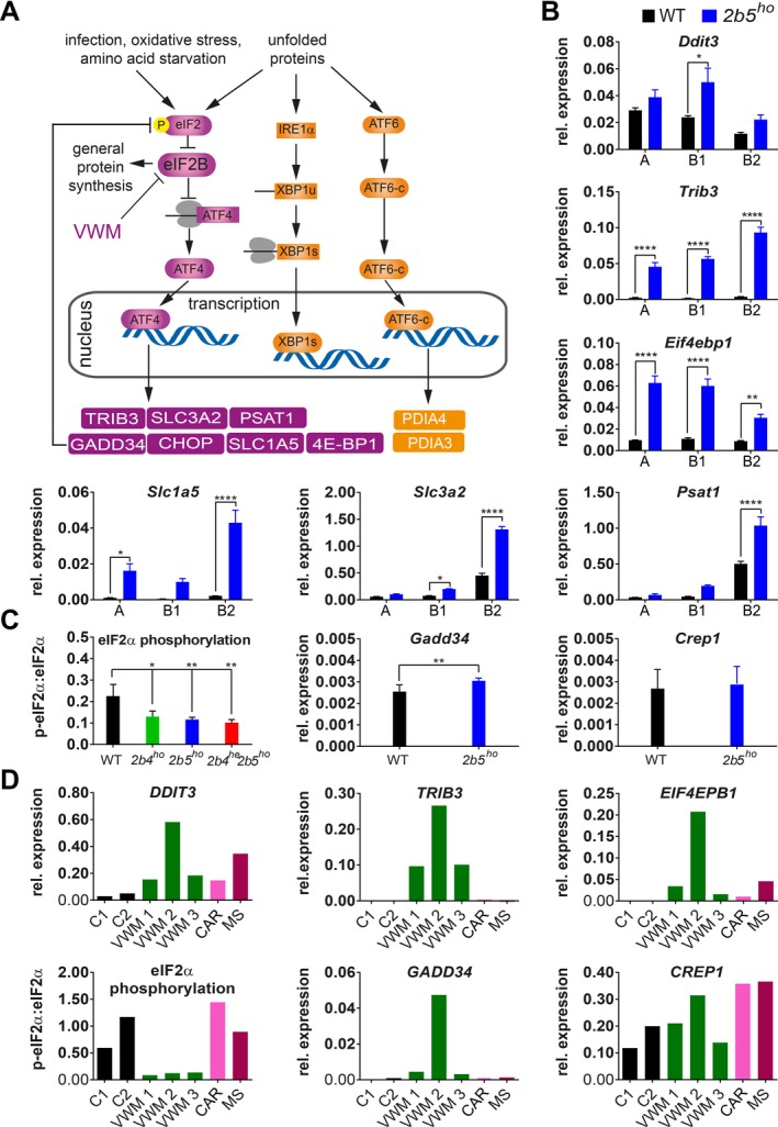
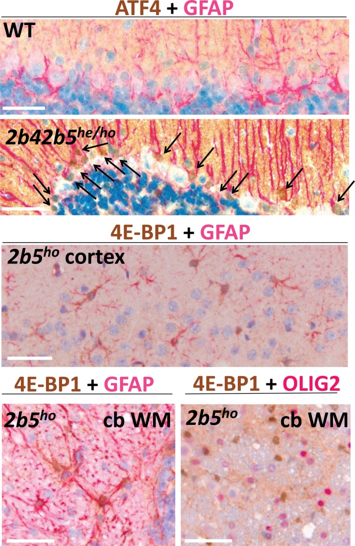
Objective: Vanishing white matter (VWM) is a fatal, stress-sensitive leukodystrophy that mainly affects children and is currently without treatment. VWM is caused by recessive mutations in eukaryotic initiation factor 2B (eIF2B) that is crucial for initiation of mRNA translation and its regulation during the integrated stress response (ISR). Mutations reduce eIF2B activity. VWM pathomechanisms remain unclear. In contrast with the housekeeping function of eIF2B, astrocytes are selectively affected in VWM. One study objective was to test our hypothesis that in the brain translation of specific mRNAs is altered by eIF2B mutations, impacting primarily astrocytes. The second objective was to investigate whether modulation of eIF2B activity could ameliorate this altered translation and improve the disease.

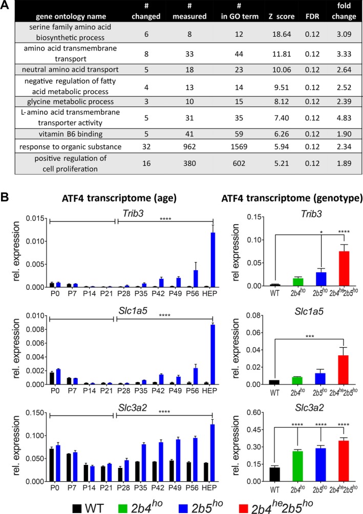
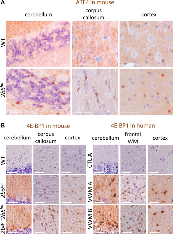
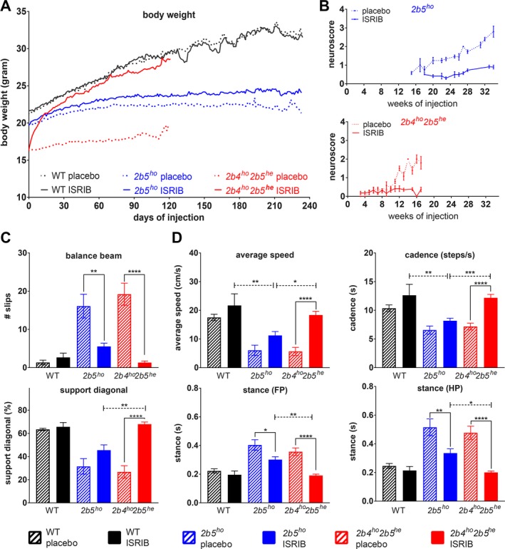
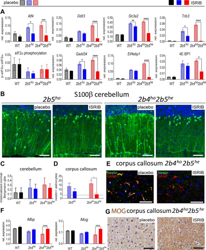
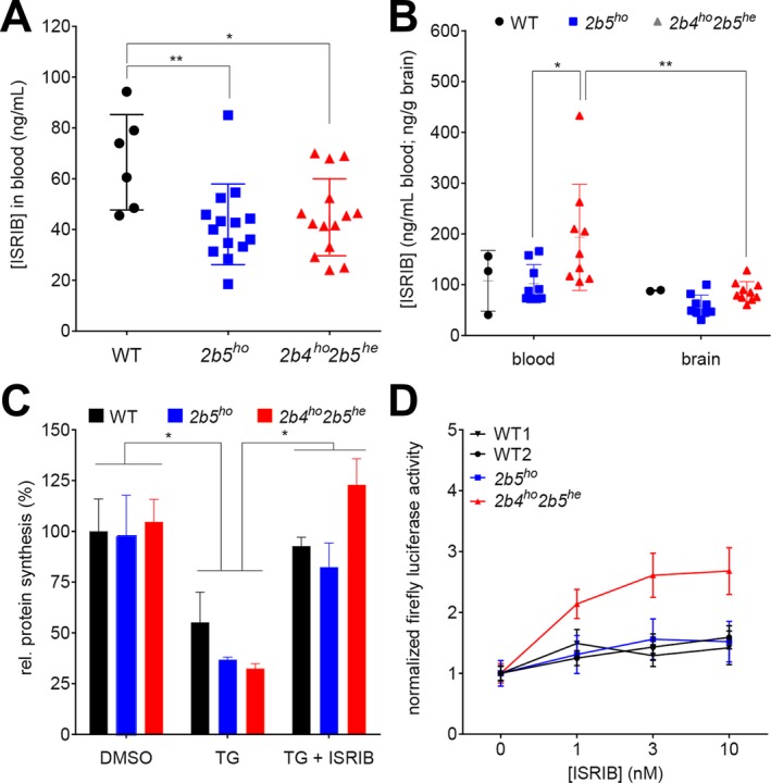
Methods: Mice with biallelic missense mutations in eIF2B that recapitulate human VWM were used to screen for mRNAs with altered translation in brain using polysomal profiling. Findings were verified in brain tissue from VWM patients using qPCR and immunohistochemistry. The compound ISRIB (for "ISR inhibitor") was administered to VWM mice to increase eIF2B activity. Its effect on translation, neuropathology, and clinical signs was assessed.

Results: In brains of VWM compared to wild-type mice we observed the most prominent changes in translation concerning ISR mRNAs; their expression levels correlated with disease severity. We substantiated these findings in VWM patients' brains. ISRIB normalized expression of mRNA markers, ameliorated brain white matter pathology and improved motor skills in VWM mice.

Interpretation: The present findings show that ISR deregulation is central in VWM pathomechanisms and a viable target for therapy.


Conflict of interest statement

MSvdK and TEMA have a patent PCT/NL2018/050293 on Guanabenz in VWM pending. Otherwise, the authors have declared that no competing interest exists.

Figures

Figure 1
Figure 1
Polysomal profiling of 2b5ho mouse brain identifies ISR deregulation in mouse VWM and human VWM brain. (A) Summary of ISR‐ and UPR‐regulated transcription included to clarify the link between the ATF4, ATF6‐c, and XBP1s transcription factors and mRNA targets investigated in this study. (B) ATF4‐regulated mRNA levels in gradient fractions from forebrain lysates from 4‐month‐old WT and 2b5ho mice were measured to visualize the mRNA distribution in monosome fraction (A), polysome fractions (B1, less than 5 ribosomes per mRNA; B2, 5 or more ribosomes per mRNA). Graphs show average ± SD, n = 3 (Akt, qPCR reference). Statistical significance was determined by two‐way ANOVA with Sidak’s correction; *P < 0.05, **P < 0.01, ****P < 0.0001). Raw data for reference mRNAs are shown in Data S7. (C) eIF2α phosphorylation, Gadd34 and Crep1 mRNA expression levels were measured in cerebellar tissue from WT and VWM mice, as indicated. Graphs show average ± SD, n = 3 for eIF2α phosphorylation and n = 6 for mRNA expression (Gapdh + Akt, qPCR reference). Statistical differences in eIF2α phosphorylation were determined using a one‐way ANOVA followed by a Dunnet’s correction. Differences between WT and 2b5ho qPCR data were assessed with Student’s t‐test, *P < 0.05, **P < 0.01. (D) DDIT3, TRIB3, EIF4EBP1 mRNA levels, eIF2α phosphorylation, GADD34 and CREP1 mRNA levels were quantified in postmortem frontal white matter tissue from VWM patients and controls (negative controls without brain pathology, C1, C2, and disease controls CAR and MS; CAR, CARASAL; MS, multiple sclerosis). Expression differences among VWM patients inversely correlate with postmortem delay time (AKT + GAPDH, qPCR reference, n = 1). Details on control and patients’ tissue are listed in Data S5. Statistical test outcomes are in Data S6.
Figure 2
Figure 2
Follow‐up analyses show affected cellular functions and ISR deregulation correlating with disease development. (A) Overrepresentation analysis of regulated polysome‐associated mRNAs in 2b5ho mouse forebrain against the gene ontology (GO) database. # changed, number of mRNAs in GO term differentially associated with 2b5ho polysomes; # measured, number of mRNAs in GO term detected in WT and 2b5ho polysomes; # in ontology, total gene number in GO term; results with a Z score >2 and FDR of <0.15 were considered significant; fold change, average of differentially expressed genes within the dataset for the particular GO term. (B) Expression of the ATF4‐regulated transcriptome in VWM mouse brain correlates positively with disease development. mRNA levels were determined at indicated ages (left hand side) or in indicated genotypes (right hand side). Sagittally sliced brain halves of WT and 2b5ho mice were analyzed at indicated ages (Gapdh, qPCR reference; P, postnatal day; HEP, humane end point, as defined by weight loss of more than 15% of body weight for 2 consecutive days and severe ataxia; 7–8 months of age). Graphs show average ± SD, n = 2. Statistical analysis on the differences at groups of different ages was done for each mRNA using a two‐way ANOVA (genotype*day interaction all days) followed by 2 two‐way ANOVAs for each mRNA (one comparing data of <P21 with ≥P21 and one comparing data of <P28 with ≥P28 for genotype*day interaction) using SPSS. Cerebella of indicated mouse genotypes were analyzed at 4‐month‐old age (Gapdh + Akt, qPCR reference). Graphs show average ± SD, n = 3 (n = 4 for 2b4ho). Statistical differences were determined using a one‐way ANOVA followed by Tukey’s correction. **P < 0.01, ***P < 0.001, ****P < 0.0001 (Data S6).
Figure 3
Figure 3
White and gray matter astrocytes in VWM mice and VWM patients are immunoreactive for ATF4‐regulated 4E‐BP1. (A) ATF4 immunoreactive nuclei (brown) are detected in white and gray matter macroglia of VWM mice. Two sections from brain tissue from 4‐month‐old WT and 2b5ho mice (n = 2) were stained with antibodies against ATF4. Findings in cerebellum, corpus callosum, and cortex are indicated. Staining for ATF4 on human brain sections was not successful. (B) White and gray matter astrocytes in VWM mouse and VWM human brain in show 4E‐BP1 immunoreactivity (brown). Two brain sections from 4‐month‐old mice (WT, 2b5ho, and 2b4he2b5ho), human control (C1) or patients (VWM 1 and VWM 2) were stained with antibodies against 4E‐BP1. Details on human brain tissue are listed in Data S5. Findings in cerebellum, corpus callosum (mouse) or frontal white matter (WM, human) and cortex are shown. Purple stain indicates nuclei. White bar, 0.05 mm.
Figure 4
Figure 4
Costaining for ATF4 or 4E‐BP1 with GFAP or OLIG2 demonstrate double positive cells of astrocyte but not oligodendrocyte lineage in VWM mice. ATF4‐GFAP costaining was only successful on sections from 2b4he2b5ho mice (4‐month‐old). Arrows indicate nuclei of double positive Bergmann glia. Shown are representative sections from a total of four sections (n = 2 animals per group). 4E‐BP1 + GFAP staining of astrocytes in cortex is at 2‐month‐old age. 4E‐BP1 + GFAP and 4E‐BP1 + OLIG2 double staining of cerebellar white matter (cb WM) is at an age of 12 months (HEP, 2b5ho). Double positive astrocytes were prominent, whereas oligodendrocytes were not detected, even at HEP. Shown are representative sections from a total of 3–4 sections (n = 2 animals per group).
Figure 5
Figure 5
ISRIB ameliorates clinical signs in two VWM mouse models, most effectively in 2b4ho2b5he mice. Mice were injected daily with vehicle or 1 mg/kg ISRIB from an age of 6–8 weeks onwards. (A–D) Graphs show phenotypic measures of placebo‐ and ISRIB‐treated WT (n = 6 per condition) and VWM mice (2b5ho and 2b4ho2b5he n = 14 per condition or as indicated): average body weight of WT and VWM mice (A), average neuroscore in VWM mice (B), average number of slips on balance beam (2b5ho n = 11 for placebo, n = 13 for ISRIB and 2b4ho2b5he n = 10 for placebo and n = 13 for ISRIB, (C), average measures of selected CatWalk parameters (2b5ho n = 9 for placebo, n = 13 for ISRIB and 2b4ho2b5he n = 12 for placebo and n = 14 for ISRIB, (D). Error bars indicate SD (graph b), SEM (graphs in c, d), or are left out (graph in a) to allow visualization of the mean values (SEM is shown in Data S6). Raw data of all CatWalk parameters are given in Data S6. Statistical analysis investigating the ISRIB differences in WT, 2b5ho and 2b4ho2b5he was performed with a two‐way ANOVA with Tukey’s correction (Data S6). *P < 0.05, **P < 0.01, ***P < 0.001, ****P < 0.0001.
Figure 6
Figure 6
ISRIB modulates aberrant expression of ATF4‐regulated mRNAs and ameliorates neuropathological astrocyte markers in VWM mouse cerebellum and corpus callosum. (A) Cerebellar expression of ATF4‐regulated mRNAs, eIF2α phosphorylation, and 4E‐BP1 protein expression were measured in placebo‐ and ISRIB‐treated WT and VWM mice (n = 6 per group). Graphs show average ± SD. Two‐way ANOVA with Tukey’s correction was performed for each target. (B) Brain sections from placebo‐ and ISRIB‐treated 2b5ho and 2b4ho2b5he mice (n = 2 per group, 2 sections per animal) were stained with antibodies against S100β. Thickness of Bergmann glia (BG) processes is reduced by ISRIB in 2b4ho2b5he mice; white bar, 0.05 mm. (C) Counts of mislocalized and normally localized S100β‐positive Bergmann glia shows that ISRIB does not fully normalize Bergmann glia location in VWM mice. Differences in the ratio of mislocalized:normal localized Bergmann glia were statistically assessed by two‐way ANOVA with Tukey’s correction. (D and E) ISRIB reduces number of nestin‐GFAP double positive astrocytes in 2b4ho2b5he mice (2 sections per rostrum and splenium of the corpus callosum for 2 animals per group). The average number of nestin‐GFAP double positive astrocytes in four untreated WT animals was included as reference (indicated as dotted line in the graph). Statistical differences were determined with one‐way ANOVA using Tukey’s correction. White bar in immunofluorescence, 0.05 mm. (F) ISRIB restored levels of mature myelin mRNA markers in 2b4ho2b5he mice. Graphs show average ± SD (Akt + Gapdh, qPCR reference). Two‐way ANOVA was performed for each target using Tukey’s correction. (G) ISRIB restores immunoreactivity for mature myelin protein MOG in white matter structures in 2b4ho2b5he mice. Shown are representative sections from a total of four sections (n = 2 mice per group). Black bar, 0.05 mm. For all graphs, *P < 0.05, **P < 0.01, ***P < 0.001, ****P < 0.0001. Statistical test outcomes are in Data S6.
Figure 7
Figure 7
ISRIB’s efficacy in VWM is influenced by the identity of eIF2B mutation. (A) ISRIB concentrations were determined in venous blood samples obtained from WT (n = 6 per group) and VWM mice (2b5ho and 2b4ho2b5he n = 14 per group) after 14 days of daily ISRIB injections (24 h after the latest injection); data obtained from placebo‐treated animals were below detection and were omitted from the graph. Statistical differences among genotypes were determined with one‐way ANOVA followed by Tukey’s correction. (B) ISRIB concentrations were determined in postmortem blood and olfactory bulb tissue from WT (n = 2 per group) and VWM mice (2b5ho and 2b4ho2b5he n = 9 and n = 8 per group). Statistical differences were determined with Two‐way ANOVA (tissue and genotype as factors) with Tukey’s correction. (C) ISRIB restored protein synthesis rates in primary cultures of adult mouse astrocytes undergoing UPR, most effectively in cells from 2b4ho2b5he mice. TG effects were not statistically significant as before29. Statistical differences in test conditions (DMSO vs. TG vs. TG + ISRIB within WT, 2b5ho, and 2b4ho2b5he adult mouse astrocytes were determined with two‐way ANOVA with Tukey’s correction (n = 2). (D) ISRIB enhanced expression of Gapdh‐driven firefly luciferase reporter in 2b4ho2b5he astrocytes at lower concentrations than in WT or 2b5ho astrocytes (P = 0.0026). WT1 and WT2 indicate different isolation dates. Cells were exposed to 300 nmol/L thapsigargin for 16 h with increasing concentrations of ISRIB. Areas under curve (AUC) were calculated for each cell isolate per transfection experiment after correcting for differences between experiments but not between test conditions within experiments (genotype, treatments). Statistical differences between ISRIB sensitivity in WT1, WT2, 2b5ho, and 2b4ho2b5he adult mouse astrocytes were determined with one‐way ANOVA with Sidak’s correction using AUC values. For all panels, *P < 0.05, **P < 0.01. Statistical test outcomes are in Data S6. Graphs show mean ± SD (A–C) or mean ± SEM (D).

References

    1. Hamilton E, van der Lei H, Vermeulen G, et al. The natural history of Vanishing White Matter. Ann Neurol 2018;84:274–288. - PMC - PubMed
    1. Bugiani M, Boor I, van Kollenburg B, et al. Defective glial maturation in vanishing white matter disease. J Neuropathol Exp Neurol 2011;70:69–82. - PMC - PubMed
    1. Li W, Wang X, Van Der Knaap MS, Proud CG. Mutations linked to leukoencephalopathy with vanishing white matter impair the function of the eukaryotic initiation factor 2B complex in diverse ways. Mol Cell Biol 2004;24:3295–3306. - PMC - PubMed
    1. Liu R, van der Lei HD, Wang X, et al. Severity of vanishing white matter disease does not correlate with deficits in eIF2B activity or the integrity of eIF2B complexes. Hum Mutat 2011;32:1036–1045. - PubMed
    1. Leegwater PA, Vermeulen G, Konst AA, et al. Subunits of the translation initiation factor eIF2B are mutant in leukoencephalopathy with vanishing white matter. Nat Genet 2001;29:383–388. - PubMed

Publication types

MeSH terms

Substances

LinkOut - more resources