

Physical Exercise and Mitochondrial Disease: Insights From a Mouse Model
- PMID: 31402893
- PMCID: PMC6673140
- DOI: 10.3389/fneur.2019.00790
Physical Exercise and Mitochondrial Disease: Insights From a Mouse Model
Abstract
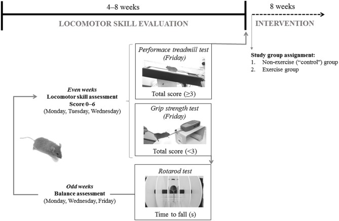
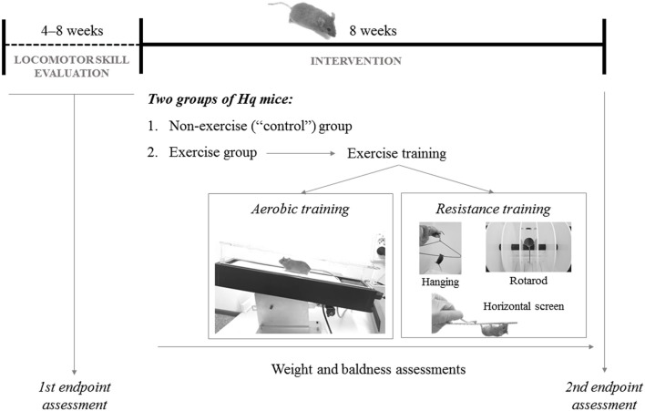
Purpose: Mitochondrial diseases (MD) are among the most prevalent neuromuscular disorders. Unfortunately, no curative treatment is yet available. This study analyzed the effects of exercise training in an animal model of respiratory chain complex I deficiency, the Harlequin (Hq) mouse, which replicates the clinical features of this condition. Methods: Male heterozygous Harlequin (Hq/Y) mice were assigned to an "exercise" (n = 10) or a "sedentary" control group (n = 11), with the former being submitted to an 8 week combined exercise training intervention (aerobic + resistance training performed five times/week). Aerobic fitness, grip strength, and balance were assessed at the beginning and at the end of the intervention period in all the Hq mice. Muscle biochemical analyses (with results expressed as percentage of reference data from age/sex-matched sedentary wild-type mice [n = 12]) were performed at the end of the aforementioned period for the assessment of major molecular signaling pathways involved in muscle anabolism (mTOR activation) and mitochondrial biogenesis (proliferator activated receptor gamma co-activator 1α [PGC-1α] levels), and enzyme activity and levels of respiratory chain complexes, and antioxidant enzyme levels. Results: Exercise training resulted in significant improvements in aerobic fitness (-33 ± 13 m and 83 ± 43 m for the difference post- vs. pre-intervention in total distance covered in the treadmill tests in control and exercise group, respectively, p = 0.014) and muscle strength (2 ± 4 g vs. 17 ± 6 g for the difference post vs. pre-intervention, p = 0.037) compared to the control group. Higher levels of ribosomal protein S6 kinase beta-1 phosphorylated at threonine 389 (156 ± 30% vs. 249 ± 30%, p = 0.028) and PGC-1α (82 ± 7% vs. 126 ± 19% p = 0.032) were observed in the exercise-trained mice compared with the control group. A higher activity of respiratory chain complexes I (75 ± 4% vs. 95 ± 6%, p = 0.019), III (79 ± 5% vs. 97 ± 4%, p = 0.031), and V (77 ± 9% vs. 105 ± 9%, p = 0.024) was also found with exercise training. Exercised mice presented with lower catalase levels (204 ± 22% vs. 141 ± 23%, p = 0.036). Conclusion: In a mouse model of MD, a training intervention combining aerobic and resistance exercise increased aerobic fitness and muscle strength, and mild improvements were found for activated signaling pathways involved in muscle mitochondrial biogenesis and anabolism, OXPHOS complex activity, and redox status in muscle tissue.
Keywords: AIF deficiency; OXPHOS; harlequin mutant mouse; mitochondrial diseases; rare diseases; resistance training; respiratory chain complex I.
Figures
Similar articles
-
Exercise Training and Neurodegeneration in Mitochondrial Disorders: Insights From the Harlequin Mouse.Front Physiol. 2020 Dec 8;11:594223. doi: 10.3389/fphys.2020.594223. eCollection 2020. Front Physiol. 2020. PMID: 33363476 Free PMC article.
-
Exercise training attenuates aging-associated mitochondrial dysfunction in rat skeletal muscle: role of PGC-1α.Exp Gerontol. 2013 Nov;48(11):1343-50. doi: 10.1016/j.exger.2013.08.004. Epub 2013 Aug 30. Exp Gerontol. 2013. PMID: 23994518
-
Thyroid hormone activation by type 2 deiodinase mediates exercise-induced peroxisome proliferator-activated receptor-γ coactivator-1α expression in skeletal muscle.J Physiol. 2016 Sep 15;594(18):5255-69. doi: 10.1113/JP272440. Epub 2016 Aug 18. J Physiol. 2016. PMID: 27302464 Free PMC article.
-
Exercise training in Tgαq*44 mice during the progression of chronic heart failure: cardiac vs. peripheral (soleus muscle) impairments to oxidative metabolism.J Appl Physiol (1985). 2017 Aug 1;123(2):326-336. doi: 10.1152/japplphysiol.00342.2017. Epub 2017 May 18. J Appl Physiol (1985). 2017. PMID: 28522765
-
Adaptation of Skeletal Muscles to Contractile Activity of Varying Duration and Intensity: The Role of PGC-1α.Biochemistry (Mosc). 2018 Jun;83(6):613-628. doi: 10.1134/S0006297918060019. Biochemistry (Mosc). 2018. PMID: 30195320 Review.
Cited by
-
Apoptosis-Inducing Factor Deficiency Induces Tissue-Specific Alterations in Autophagy: Insights from a Preclinical Model of Mitochondrial Disease and Exercise Training Effects.Antioxidants (Basel). 2022 Mar 7;11(3):510. doi: 10.3390/antiox11030510. Antioxidants (Basel). 2022. PMID: 35326160 Free PMC article.
-
Recent advancements in the understanding of the alterations in mitochondrial biogenesis in Alzheimer's disease.Mol Biol Rep. 2025 Jan 29;52(1):173. doi: 10.1007/s11033-025-10297-6. Mol Biol Rep. 2025. PMID: 39880979 Review.
-
Therapeutic Approaches to Treat Mitochondrial Diseases: "One-Size-Fits-All" and "Precision Medicine" Strategies.Pharmaceutics. 2020 Nov 11;12(11):1083. doi: 10.3390/pharmaceutics12111083. Pharmaceutics. 2020. PMID: 33187380 Free PMC article. Review.
-
Molecular Basis for the Therapeutic Effects of Exercise on Mitochondrial Defects.Front Physiol. 2021 Jan 13;11:615038. doi: 10.3389/fphys.2020.615038. eCollection 2020. Front Physiol. 2021. PMID: 33584337 Free PMC article. Review.
-
Mitochondrial Dysfunction and Oxidative Stress in Alzheimer's Disease.Front Aging Neurosci. 2021 Feb 18;13:617588. doi: 10.3389/fnagi.2021.617588. eCollection 2021. Front Aging Neurosci. 2021. PMID: 33679375 Free PMC article. Review.
References
-
- Chinnery PF. Mitochondrial disorders overview. genereviews®. In: Adam MP, Ardinger HH, Pagon RA, Wallace SE, Bean LJH, Stephens K, Amemiya A, editors. GeneReviews®. Seattle,WA: University of Washington; (2000). pp. 1993–2019.
LinkOut - more resources
Full Text Sources
Molecular Biology Databases
Miscellaneous