Review
. 2019 Jul 25:10:652.
doi: 10.3389/fgene.2019.00652. eCollection 2019.

Omics Driven Understanding of the Intestines of Parasitic Nematodes

Affiliations
Review

Omics Driven Understanding of the Intestines of Parasitic Nematodes

Douglas P Jasmer et al. Front Genet. .

Abstract

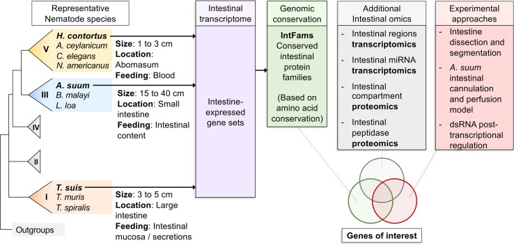
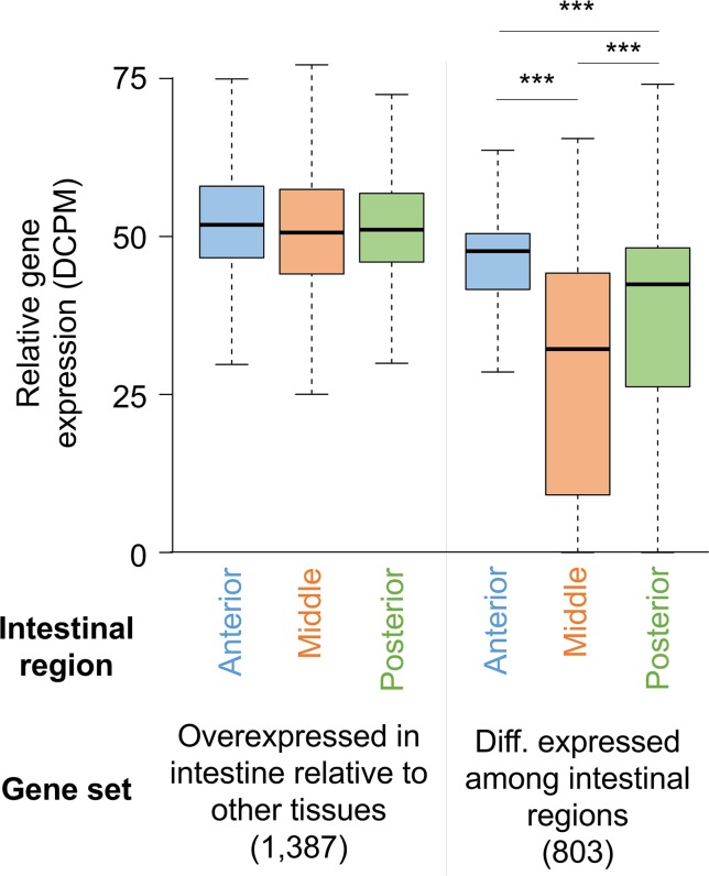
The biological and molecular complexity of nematodes has impeded research on development of new therapies for treatment and control. We have focused on the versatility of the nematode intestine as a target for new therapies. To that end, it is desirable to establish a broad and deep understanding of the molecular architecture underlying intestinal cell functions at the pan-Nematoda level. Multiomics data were generated to uncover the evolutionary principles underlying both conserved and adaptable features of the nematode intestine. Whole genomes were used to reveal the functional potential of the nematodes, tissue-specific transcriptomes provided a deep assessment of genes that are expressed in the adult nematode intestine, and comparison of selected core species was used to determine a first approximation of the pan-Nematoda intestinal transcriptome. Differentially expressed transcripts were also identified among intestinal regions, with the largest number expressed at significantly higher levels in the anterior region, identifying this region as the most functionally unique compared to middle and posterior regions. Profiling intestinal miRNAs targeting these genes identified the conserved intestinal miRNAs. Proteomics of intestinal cell compartments assigned proteins to several different intestinal cell compartments (intestinal tissue, the integral and peripheral intestinal membranes, and the intestinal lumen). Finally, advanced bioinformatic approaches were used to predict intestinal cell functional categories of seminal importance to parasite survival, which can now be experimentally tested and validated. The data provide the most comprehensive compilation of constitutively and differentially expressed genes, predicted gene regulators, and proteins of the nematode intestine. The information provides knowledge that is essential to understand molecular features of nematode intestinal cells and functions of fundamental importance to the intestine of many, if not all, parasitic nematodes.

Keywords: dsRNA; genome; intestine; miRNA; nematode; proteome; transcriptome.


Figures

Figure 1
Figure 1
Overall workflow of reviewed intestinal experiments. Transcriptomic and genomic data for core intestinal nematode species are utilized to define intestinal families. Results from additional experiments are intersected and compared to produce prioritized genes of interest to suit experimental needs.
Figure 2
Figure 2
Anatomy of model intestinal nematode species Ascaris suum. (A) Major organs are identified in an adult A. suum female worm, including (B) in cross-section.
Figure 3
Figure 3
A detailed example of one bioinformatic workflow. Here, a list of conserved intestinal-expressed protein families is produced by intersecting several datasets. The workflow for producing the Ascaris suum genome assembly and annotation is shown but omitted for the other two core species.
Figure 4
Figure 4
Distribution of gene expression levels for A. suum genes in the anterior, middle, and posterior intestine regions among all intestine-overexpressed genes and genes differentially expressed between the regions. ***P < 10−5.
Figure 5
Figure 5
The A. suum intestinal cannulation and perfusion system. Shown are (A) a diagram description of the system and (B) a picture of the actual worms set up in the system, and contained inside of plastic test tubes.
Figure 6
Figure 6
Proteomics-based inference of A. suum intestinal proteins in different compartments. (A) Anatomy of model intestinal nematode species Ascaris suum (transverse section). (B) Protein sets detected by MS/MS proteomics from samples harvested from adult A. suum worms (left) have other protein sets strategically removed (center) to deduce final protein sets in different intestinal compartments (right). “Integral intestinal membrane” proteins are not labelled as “basal” because they may include some proteins from the apical intestinal membrane as well. *Proteins annotated with “cellular compartment” Gene Ontology terms for endoplasmic reticulum, mitochondria, Golgi apparatus, and nucleus were removed to reduce contamination from proteins embedded in these organelles rather than the external cellular membrane. **Only proteins annotated with predicted classical or nonclassical secretion signals were included since these are better candidates for proteins that are transported to the membrane.

References

    1. Allman E., Johnson D., Nehrke K. (2009). Loss of the apical V-ATPase a-subunit VHA-6 prevents acidification of the intestinal lumen during a rhythmic behavior in C. elegans. Am. J. Physiol. Cell Physiol. 297 (5), 9. 10.1152/ajpcell.00284.2009 - DOI - PMC - PubMed
    1. Andrews S. J., Hole N. J., Munn E. A., Rolph T. P. (1995). Vaccination of sheep against haemonchosis with H11, a gut membrane-derived protective antigen from the adult parasite: prevention of the periparturient rise and colostral transfer of protective immunity. Int. J. Parasitol. 25 (7), 839–846. 10.1016/0020-7519(94)00221-9 - DOI - PubMed
    1. Anisimov A. P., Tokmakova N. P., Usheva L. N. (1975). Proliferation and growth of intestinal epithelium in Ascaris suum (Nematoda) during postnatal ontogeny. Communication III. Mitotic anomalies and changes in ploidy and nucleus size. Sov. J. Dev. Biol. 5 (1), 37–44. - PubMed
    1. Ashburner M., Ball C. A., Blake J. A., Botstein D., Butler H., Cherry J. M., et al. (2000). Gene ontology: tool for the unification of biology. the gene ontology consortium. Nat. Genet. 25 (1), 25–29. 10.1038/75556 - DOI - PMC - PubMed
    1. Atkinson C., Newsam R. J., Gull K. (1980). Influence of the antimicrotubule agent, mebendazole, on the secretory activity of intestinal cells of Ascaridia galli. Protoplasma 105 (1-2), 69–76. 10.1007/BF01279850 - DOI - PubMed

LinkOut - more resources