. 2019 Aug;6(4):241-247.
doi: 10.1093/rb/rbz009. Epub 2019 Mar 18.

Bone remodeling effect of a chitosan and calcium phosphate-based composite

Affiliations

Bone remodeling effect of a chitosan and calcium phosphate-based composite

Lilja Kjalarsdóttir et al. Regen Biomater. 2019 Aug.

Abstract

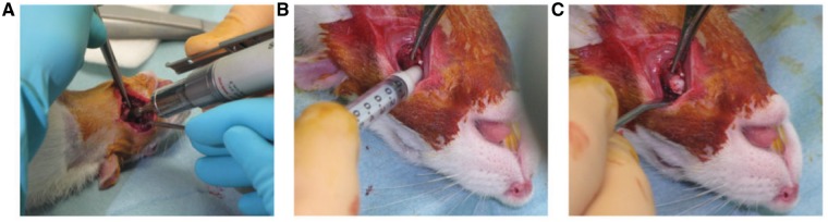
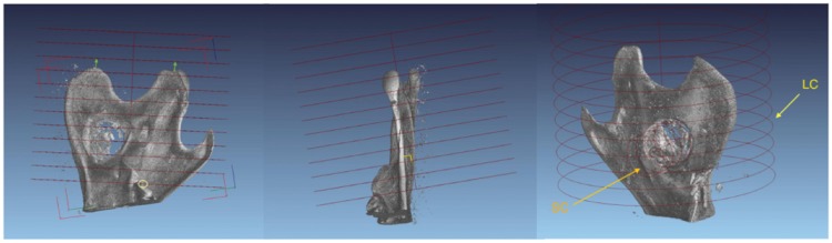
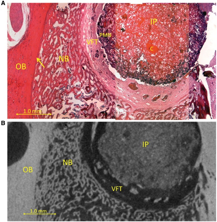
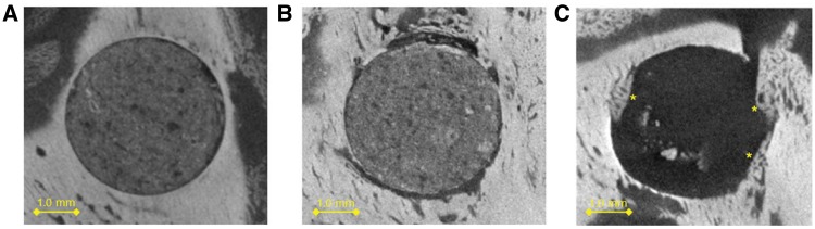
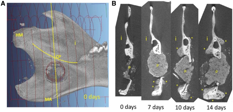
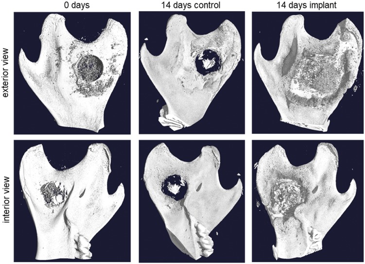
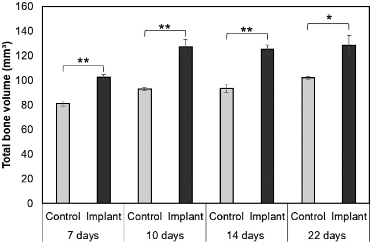
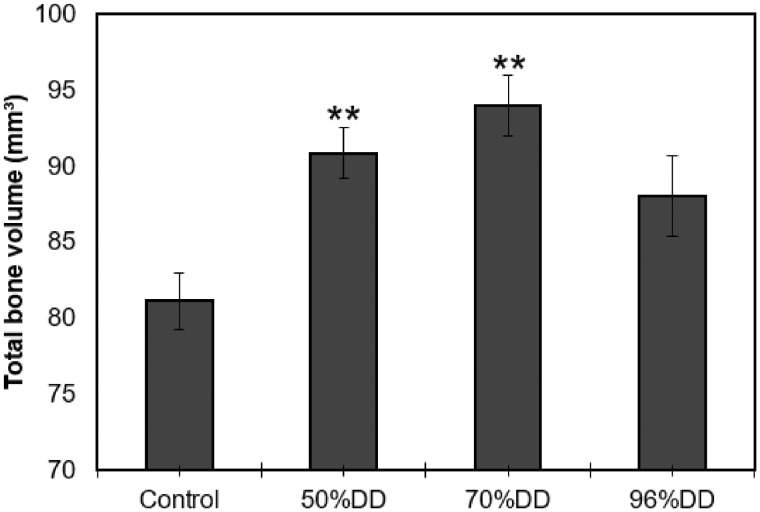
Chitosan is a biocompatible polymer that has been widely studied for tissue engineering purposes. The aim of this research was to assess bone regenerative properties of an injectable chitosan and calcium phosphate-based composite and identify optimal degree of deacetylation (%DDA) of the chitosan polymer. Drill holes were generated on the left side of a mandible in Sprague-Dawley rats, and the hole was either left empty or filled with the implant. The animals were sacrificed at several time points after surgery (7-22 days) and bone was investigated using micro-CT and histology. No significant new bone formation was observed in the implants themselves at any time points. However, substantial new bone formation was observed in the rat mandible further away from the drill hole. Morphological changes indicating bone formation were found in specimens explanted on Day 7 in animals that received implant. Similar bone formation pattern was seen in control animals with an empty drill hole at later time points but not to the same extent. A second experiment was performed to examine if the %DDA of the chitosan polymer influenced the bone remodeling response. The results suggest that chitosan polymers with %DDA between 50 and 70% enhance the natural bone remodeling mechanism.

Keywords: bone defects; bone implant; bone remodeling; chitosan; degree of deacetylation; micro-CT; rat mandible.


Figures

Figure 1
Figure 1
Overview of surgery in a rat receiving implant. (A) Dental drill was used to drill the hole. (B) Implant injected into the hole. (C) Appearance of tissue after implantation showing how implant fills the hole
Figure 2
Figure 2
Standardization of a partial mandibular volume from micro-CT data according to anatomical landmarks in order to enable comparison of total bone volume. Orientation of the larger cylinder (LC) embracing the entire posterior part of the mandible. One end of the cylinder was positioned so as to sit on the innermost molar and the orientation of the cylinder was adjusted so as to be perpendicular to an imagined plane sitting on HM and the end of the angular process. The smaller cylinder (SC) also indicated covered the drillhole encompassing all remaining bone splinter as well as the implant
Figure 3
Figure 3
(A) Drilled hole with 14 days old implant (IP) containing PMN leukocytes (PMN) stained blue in the outer rim of the implant, then vascular fibrous tissue (FT), then new trabecular bone (NB). Original bone is on the left (OB) and the edge between old and new bone is indicated (arrow). Undissolved chitosan can be seen in the implant (C). (B) Virtual sectioning through a micro-CT image of the same sample. Drilled hole with implant (IP) then vascularized fibrous tissue (VFT, dark layer), then new trabecular bone (NB). Original bone (OB) is on the left
Figure 4
Figure 4
Micro-CT image sectioning of implant comparing Day 7 (A) and Day 22 (B). (C) Control sample at Day 22. No significant bone formation was observed in the implants. In the control sample, little new bone (*) formation was observed
Figure 5
Figure 5
The effect of implant on mineralized tissue formation as a function of time in the rat mandible. (A, left): Overview of the interior side of the mandible indicating the drilled hole. Yellow line shows the location of the transverse section. (B, right): Four transverse sections of 0, 7, 10 and 14 days post operation showing the implant (p) and the formation of new mineralized tissue (*). i, interior side; MA, mandibular angle; CP, condylar process; MH, head of mandible. For the 0 days time point, rats were sacrificed at the same day as the operation
Figure 6
Figure 6
Three-dimensional reconstruction from micro-CT data comparing a control sample and a sample with implanted composite with 70% DDA chitosan after 14 days of incubation. The control sample (center) shows some thickening of the bone around the hole as a natural response to the injury. The sample with implant (right) shows a much more distinct thickening of the bone in the same areas around the hole. As a reference, a sample from an animal that died in surgery is shown (left). As the implant material did not have time to set, most of it was washed away, leaving some remnants around the hole
Figure 7
Figure 7
Change in total bone volume in a defined part of rat mandible as a function of time. Results from two-tailed t-test, statistical significance between control and implant groups for each time is indicated (*P < 0.05, **P < 0.01). Graphs represent mean±SEM
Figure 8
Figure 8
Change in total bone volume in a defined part of rat mandible as a function of implant type (%DDA). Results from one-way ANOVA comparing all treatment groups to the control (**P < 0.01). Graphs represent mean±SEM

References

    1. Oryan A, Alidadi S, Moshiri A. et al. Bone regenerative medicine: classic options, novel strategies, and future directions. J Orthop Surg Res 2014;9:18.. - PMC - PubMed
    1. Dimitriou R, Jones E, McGonagle D. et al. Bone regeneration: current concepts and future directions. BMC Med 2011;9:66.. - PMC - PubMed
    1. Dennis SC, Berkland CJ, Bonewald LF. et al. Endochondral ossification for enhancing bone regeneration: converging native extracellular matrix biomaterials and developmental engineering in vivo. Tissue Eng Part B Rev 2015;21:247–66. - PMC - PubMed
    1. Sheikh Z, Javaid MA, Hamdan N. et al. Bone regeneration using bone morphogenetic proteins and various biomaterial carriers. Materials (Basel) 2015;8:1778–816. - PMC - PubMed
    1. Carragee EJ, Hurwitz EL, Weiner BK.. A critical review of recombinant human bone morphogenetic protein-2 trials in spinal surgery: emerging safety concerns and lessons learned. Spine J 2011;11:471–91. - PubMed