. 2019 Sep 1;19(6):foz057.
doi: 10.1093/femsyr/foz057.

Impact of manganese on biofilm formation and cell morphology of Candida parapsilosis clinical isolates with different biofilm forming abilities

Affiliations

Impact of manganese on biofilm formation and cell morphology of Candida parapsilosis clinical isolates with different biofilm forming abilities

Sulman Shafeeq et al. FEMS Yeast Res. .

Abstract

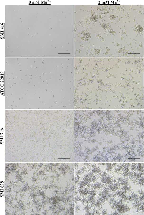
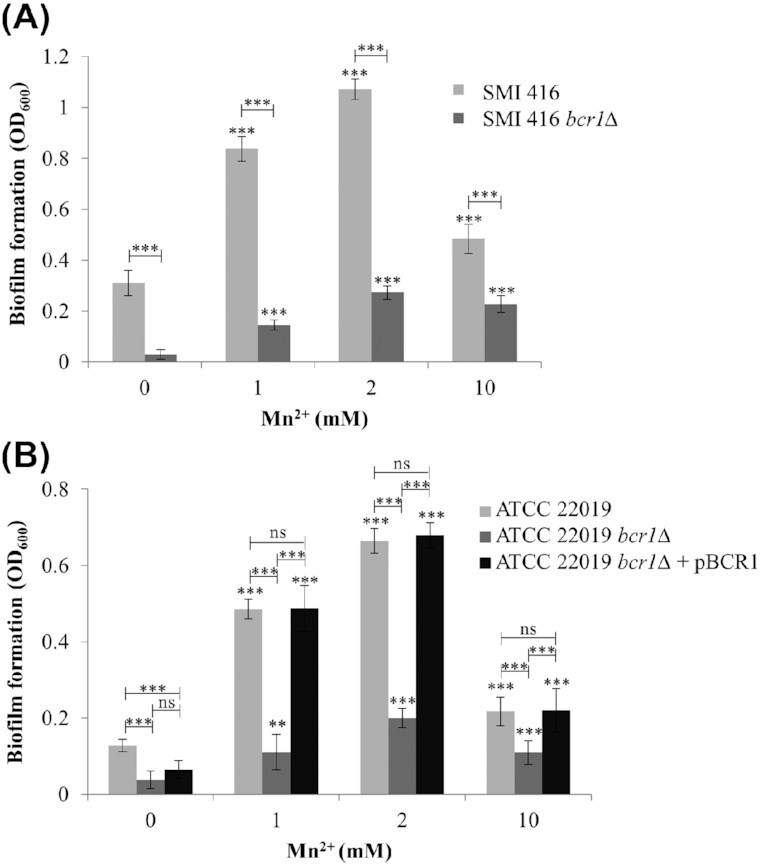
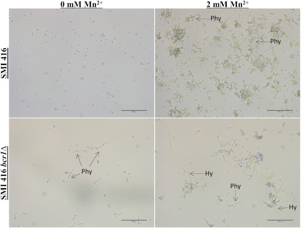
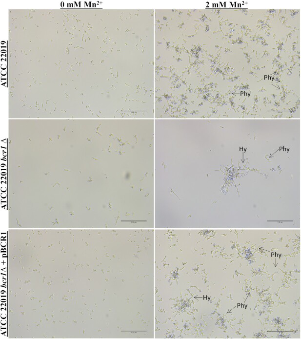
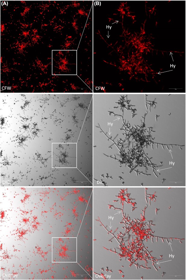
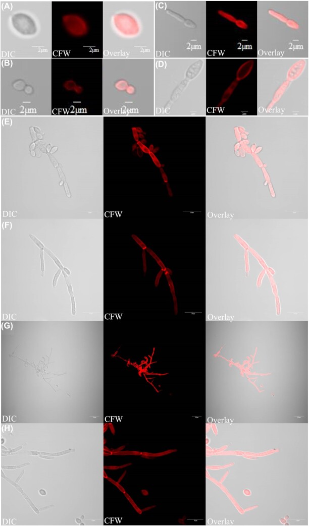
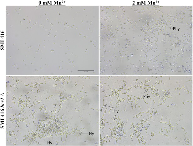
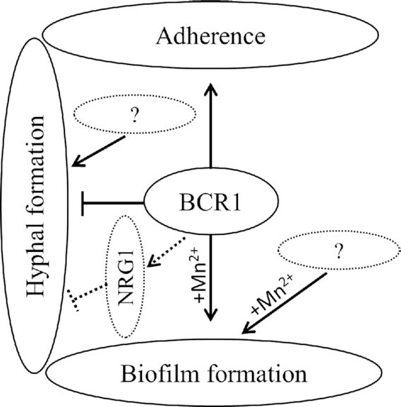
The commensal species Candida parapsilosis is an emerging human pathogen that has the ability to form biofilms. In this study, we explored the impact of the divalent cations cobalt (Co2+), copper (Cu2+), iron (Fe3+), manganese (Mn2+), nickel (Ni2+) and zinc (Zn2+) on biofilm formation of clinical isolates of C. parapsilosis with no, low and high biofilm forming abilities at 30 and 37°C. All strains besides one isolate showed a concentration-dependent enhancement of biofilm formation at 30°C in the presence of Mn2+ with a maximum at 2 mM. The biofilm forming ability of no and low biofilm forming isolates was >2-fold enhanced in the presence of 2 mM Mn2+, while the effect in high biofilm forming isolate was significantly less pronounced. Of note, cells in the biofilms of no and low biofilm forming strains differentiated into yeast and pseudohyphal cells similar in morphology to high biofilm formers. The biofilm transcriptional activator BCR1 has a dual developmental role in the absence and presence of 2 mM Mn2+ as it promoted biofilm formation of no biofilm forming strains, and, surprisingly, suppressed cells of no biofilm forming strains to develop into pseudohyphae and/or hyphae. Thus, environmental conditions can significantly affect the amount of biofilm formation and cell morphology of C. parapsilosis with Mn2+ to overcome developmental blocks to trigger biofilm formation and to partially relieve BCR1 suppressed cell differentiation.

Keywords: BCR1; Candida parapsilosis; biofilm formation; cell morphology; hyphae; manganese; metal ions; pseudohyphae; yeast cells.


Figures

Figure 1.
Figure 1.
Impact of increasing concentration of Mn2+ on the biofilm forming ability of no (SMI 416 and ATCC 22019), low (SMI 706) and high (SMI 828) biofilm formers of C. parapsilosis on a polystyrene surface in YNB/2% glucose medium after 48 h at 30°C. (A) SMI 416, (B) ATCC 22019, (C) SMI 706 and (D) SMI 828. Values are the mean of three independent experiments conducted in three biological replicates. Error bars indicate standard deviation. Statistical significance of the differences in biofilm formation in the presence of Mn2+ compared to 0 mM Mn2+ was determined by one-way ANOVA (ns—not significant, *P < 0.05, **P < 0.01 and ***P < 0.001).
Figure 2.
Figure 2.
Impact of Mn2+ on biofilm formation and cell morphology on a polystyrene surface of no (SMI 416 and ATCC 22019), low (SMI 706) and high (SMI 828) biofilm formers of C. parapsilosis at 30°C. Scale bar represents 100 μm.
Figure 3.
Figure 3.
Impact of different concentrations of Mn2+ on the biofilm forming ability of wild-types SMI 416 and ATCC 22019, bcr1Δ mutants SMI 416 bcr1Δ and ATCC 22019 bcr1Δ, and BCR1 complementation ATCC 22019 bcr1Δ + pBCR1 on a polystyrene surface in YNB/2% glucose medium at 30°C after 48 h. Values are the mean of three independent experiments conducted in three biological replicates. Error bars indicate standard deviation. Statistical significance of the differences in biofilm formation in the presence of Mn2+ compared to the absence of Mn2+ and wild-type compared to bcr1Δ mutant/BCR1 complementation was determined by one-way ANOVA (ns—not significant, **P < 0.01 and ***P < 0.001).
Figure 4.
Figure 4.
Impact of the BCR1 deletion on biofilm formation and cell morphology of wild-type C. parapsilosis SMI 416 and its bcr1Δ mutant at 30°C on a polystyrene surface in YNB/2% glucose medium with 0 and 2 mM of Mn2+. Arrows indicate pseudohyphae (Phy) and hyphae (Hy). Scale bar represents 100 μm.
Figure 5.
Figure 5.
Impact of BCR1 deletion on biofilm formation and cell morphology of C. parapsilosis ATCC 22019 wild-type, its bcr1Δ mutant and BCR1 complementation at 30°C on a polystyrene surface in YNB/2% glucose medium with 0 and 2 mM of Mn2+. Arrows indicates the presence of pseudohyphae (Phy) and hyphae (Hy). Scale bar represents 100 μm.
Figure 6.
Figure 6.
Visualization of hyphal development in the bcr1Δ mutant of C. parapsilosis SMI 416 at 30°C on a polystyrene surface in YNB/2% glucose medium with 2 mM Mn2+. Arrows indicate the presence of hyphae (Hy). Scale bars: (A) 200 μm and (B) 50 μm.
Figure 7.
Figure 7.
Different cell morphologies observed in this study: (A) blastospore, (B) budding yeast, (C, D) germ tube, (E) pseudohyphae and (F–H) hyphae. Light (left) and fluorescent (Calcofluor staining; middle) microscopy images of different cell types were overlaid (right). Scale bars: (A–D) 2 μm, (E, F, H) 10 μm and (G) 20 μm.
Figure 8.
Figure 8.
YPD/2% glucose medium induces filamentous growth of C. parapsilosis SMI 416 in a polystyrene surface. Cell morphology of C. parapsilosis SMI 416 wild-type and bcr1Δ mutant in YPD/2% glucose medium with and without 2 mM Mn2+ at 30°C. Scale bar represents 100 μm.
Figure 9.
Figure 9.
A tentative model of the role of Bcr1 in no biofilm forming isolates of C. parapsilosis.

Similar articles

Cited by

References

    1. Allison DL, Willems HM, Jayatilake JAet al. .. Candida–bacteria interactions: their impact on human disease. Microbiol Spectr. 2016;4:VMBF-0030-2016, DOI:10.1128/microbiolspec.VMBF-0030-2016. - DOI - PubMed
    1. Araújo D, Henriques M, Silva S. Portrait of Candida species biofilm regulatory network genes. Trends Microbiol. 2017;25:62–75. - PubMed
    1. Bastidas RJ, Heitman J, Cardenas ME. The protein kinase Tor1 regulates adhesin gene expression in Candida albicans. PLoS Pathog. 2009;5:e1000294. - PMC - PubMed
    1. Braun BR, Kadosh D, Johnson AD. NRG1, a repressor of filamentous growth in C. albicans, is down-regulated during filament induction. EMBO J. 2001;20:4753–61. - PMC - PubMed
    1. Cavalheiro M, Teixeira MC. Candida biofilms: threats, challenges, and promising strategies. Front Med. 2018;5:28. - PMC - PubMed

Publication types

MeSH terms