

Association of Chronic Active Multiple Sclerosis Lesions With Disability In Vivo
- PMID: 31403674
- PMCID: PMC6692692
- DOI: 10.1001/jamaneurol.2019.2399
Association of Chronic Active Multiple Sclerosis Lesions With Disability In Vivo
Erratum in
-
Errors in Table and Supplemental Figure.JAMA Neurol. 2019 Dec 1;76(12):1520. doi: 10.1001/jamaneurol.2019.4331. JAMA Neurol. 2019. PMID: 31816021 Free PMC article. No abstract available.
Abstract
Importance: In multiple sclerosis (MS), chronic active lesions, which previously could only be detected at autopsy, can now be identified on susceptibility-based magnetic resonance imaging (MRI) in vivo as non-gadolinium-enhancing lesions with paramagnetic rims. Pathologically, they feature smoldering inflammatory demyelination at the edge, remyelination failure, and axonal degeneration. To our knowledge, the prospect of long-term in vivo monitoring makes it possible for the first time to determine their contribution to disability and value as a treatment target.
Objective: To assess whether rim lesions are associated with patient disability and long-term lesion outcomes.
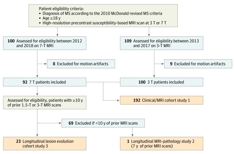
Design, setting, participants: We performed 3 studies at the National Institutes of Health Clinical Center: (1) a prospective clinical/radiological cohort of 209 patients with MS (diagnosis according to the 2010 McDonald revised MS criteria, age ≥18 years, with 7-T or 3-T susceptibility-based brain MRI results) who were enrolled from January 2012 to March 2018 (of 209, 17 patients [8%] were excluded because of uninterpretable MRI scans); (2) a radiological/pathological analysis of expanding lesions featuring rims; and (3) a retrospective longitudinal radiological study assessing long-term lesion evolution in 23 patients with MS with yearly MRI scans for 10 years or more (earliest scan, 1992).
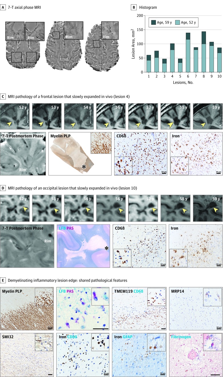
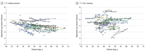
Main outcomes and measures: (1) Identification of chronic rim lesions on 7-T or 3-T susceptibility-based brain MRI in 192 patients with MS and the association of rim counts with clinical disability (primary analysis) and brain volume changes (exploratory analysis). (2) Pathological characterization of 10 expanding lesions from an adult with progressive MS who came to autopsy after 7 years of receiving serial in vivo MRI scans. (3) Evaluation of annual lesion volume change (primary analysis) and T1 times (exploratory analysis) in 27 rim lesions vs 27 rimless lesions.
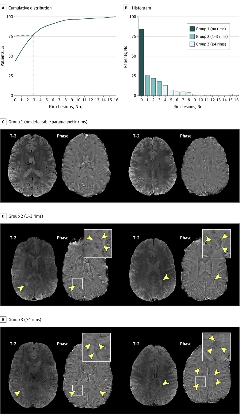
Results: Of 209 participants, 104 (50%) were women and 32 (15%) were African American. One hundred seventeen patients (56%) had at least 1 rim lesion regardless of prior or ongoing treatment. Further, 84 patients (40%) had no rims (mean [SD] age, 47 [14] years), 66 (32%) had 1 to 3 rims (mean [SD] age, 47 [11] years), and 42 (20%) had 4 rims or more (mean [SD] age, 44 [11] years). Individuals with 4 rim lesions or more reached motor and cognitive disability at an earlier age. Normalized volumes of brain, white matter, and basal ganglia were lower in those with rim lesions. Whereas rimless lesions shrank over time (-3.6%/year), rim lesions were stable in size or expanded (2.2%/year; P < .001). Rim lesions had longer T1 times, suggesting more tissue destruction, than rimless lesions. On histopathological analysis, all 10 rim lesions that expanded in vivo had chronic active inflammation.
Conclusions and relevance: Chronic active lesions are common, are associated with more aggressive disease, exert ongoing tissue damage, and occur even in individuals treated with effective disease-modifying therapies. These results prompt the planning of MRI-based clinical trials aimed at treating perilesional chronic inflammation in MS.
Conflict of interest statement
Figures
References
-
- Luchetti S, Fransen NL, van Eden CG, Ramaglia V, Mason M, Huitinga I. Progressive multiple sclerosis patients show substantial lesion activity that correlates with clinical disease severity and sex: a retrospective autopsy cohort analysis. Acta Neuropathol. 2018;135(4):511-528. doi: 10.1007/s00401-018-1818-y - DOI - PMC - PubMed
Publication types
MeSH terms
LinkOut - more resources
Full Text Sources
Other Literature Sources
Medical
