. 2019 Sep 24;13(9):9980-9992.
doi: 10.1021/acsnano.8b09137. Epub 2019 Aug 20.

Engineering and Modeling the Electrophoretic Trapping of a Single Protein Inside a Nanopore

Affiliations

Engineering and Modeling the Electrophoretic Trapping of a Single Protein Inside a Nanopore

Kherim Willems et al. ACS Nano. .

Abstract

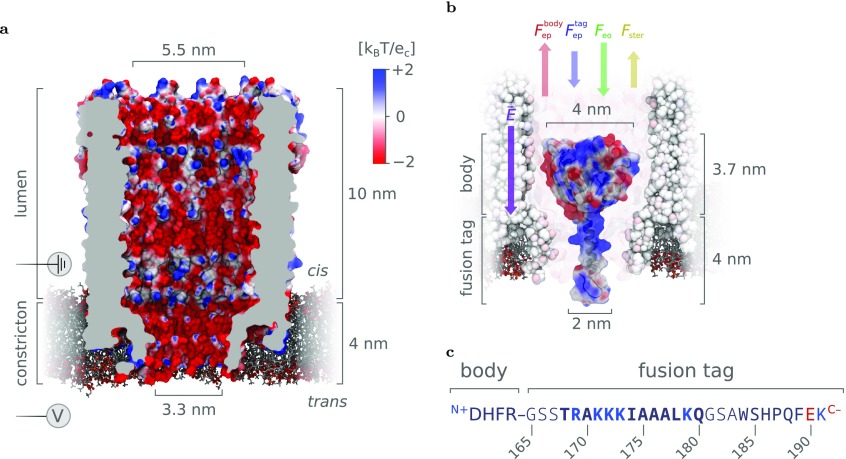
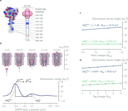
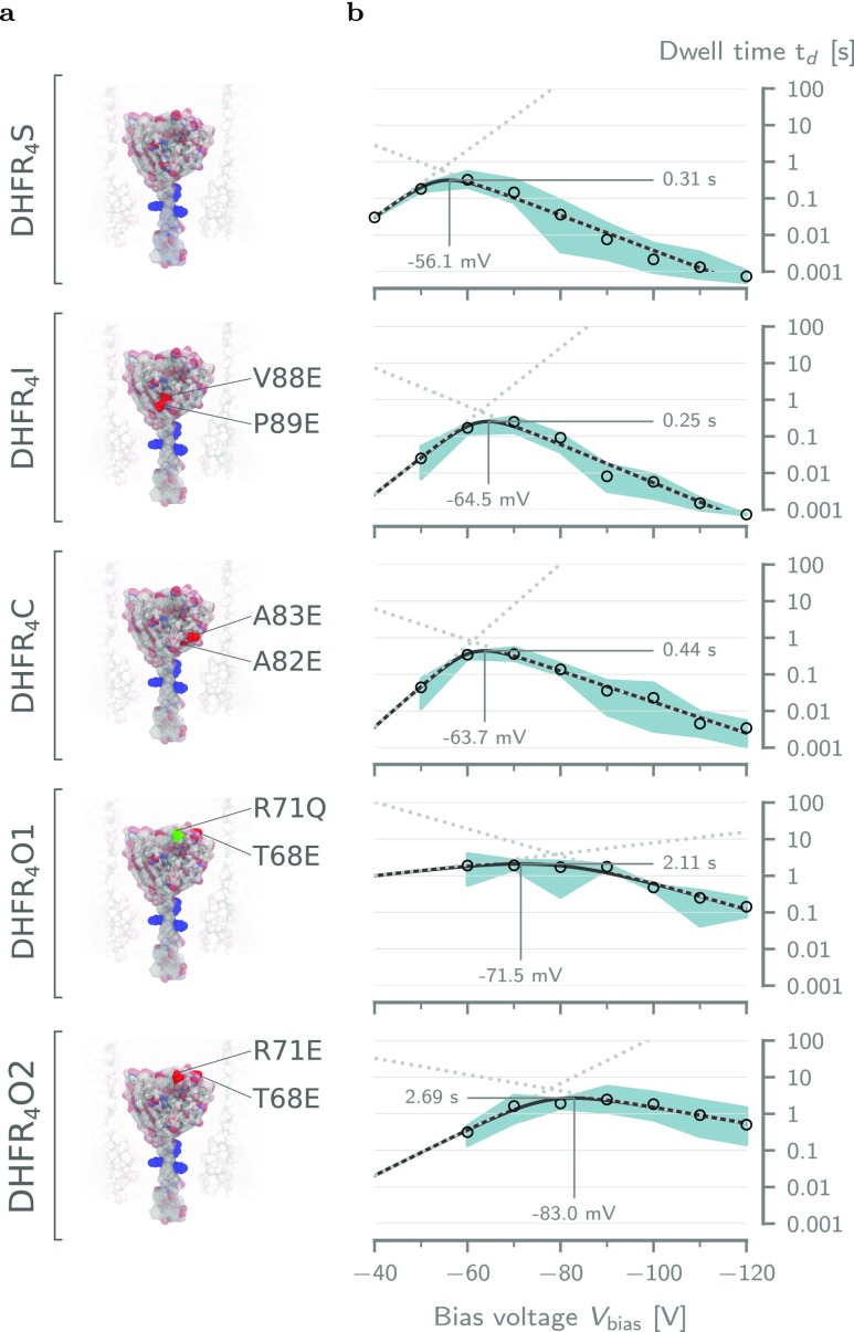
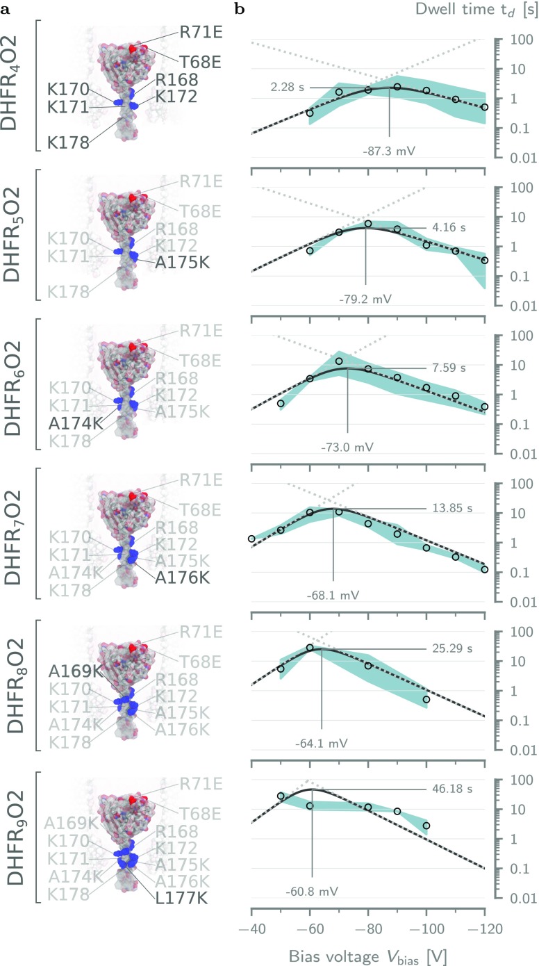
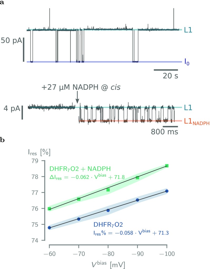
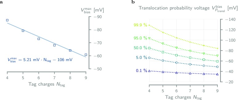
The ability to confine and to study single molecules has enabled important advances in natural and applied sciences. Recently, we have shown that unlabeled proteins can be confined inside the biological nanopore Cytolysin A (ClyA) and conformational changes monitored by ionic current recordings. However, trapping small proteins remains a challenge. Here, we describe a system where steric, electrostatic, electrophoretic, and electro-osmotic forces are exploited to immobilize a small protein, dihydrofolate reductase (DHFR), inside ClyA. Assisted by electrostatic simulations, we show that the dwell time of DHFR inside ClyA can be increased by orders of magnitude (from milliseconds to seconds) by manipulation of the DHFR charge distribution. Further, we describe a physical model that includes a double energy barrier and the main electrophoretic components for trapping DHFR inside the nanopore. Simultaneous fits to the voltage dependence of the dwell times allowed direct estimates of the cis and trans translocation probabilities, the mean dwell time, and the force exerted by the electro-osmotic flow on the protein (≅9 pN at -50 mV) to be retrieved. The observed binding of NADPH to the trapped DHFR molecules suggested that the engineered proteins remained folded and functional inside ClyA. Contact-free confinement of single proteins inside nanopores can be employed for the manipulation and localized delivery of individual proteins and will have further applications in single-molecule analyte sensing and enzymology studies.

Keywords: ClyA nanopore; DHFR; electro-osmotic flow; electrostatic trap; nanomanipulation; protein electrostatics; single-molecule enzymology.


Conflict of interest statement

The authors declare no competing financial interest.

Figures

Figure 1
Figure 1
Trapping of proteins inside the ClyA-AS nanopore. (a) Surface representation of a type I ClyA-AS nanopore—a dodecameric version of the Cytolysin A pore containing eight mutations (C87A, L99Q, E103G, F166Y, I203V, C285S, K294R, H307Y) compared to the wild-type variant from Salmonella typhimurium— embedded in a planar lipid bilayer. The structure was derived through homology modeling from the wild-type crystal structure (PDB ID: 2WCD(56)) using the MODELLER, VMD, and NAMD software packages. The surface of the pore is colored according to its electrostatic potential in 150 mM NaCl, as calculated by APBS. (b) Depiction of a single dihydrofolate reductase (DHFR) molecule extended with a positively charged C-terminal polypeptide tag (DHFR4S) inside a ClyA-AS nanopore. The secondary structure of the tag (primarily α-helical) was predicted by the PEP-FOLD server., At negative applied bias voltages relative to trans, the electric field (E⃗) is expected to pull the negatively charged body of DHFR upward (Fepbody) and the positively charged fusion tag downward (Feptag), while the electro-osmotic flow pushes the entire protein downward (Feo). Lastly, as the body of DHFR is larger than the diameter of the trans constriction, the force required to overcome the steric hindrance (Fster) during full cis-to-trans translocation is expected to be significant. (c) Sequence of DHFR4S fusion tag with its positive and negative residues colored blue and red, respectively. The sequence of the Strep-tag starts at residue 183, and the GSS and GSA linkers are shown in light font. Note that, at pH 7.5, the C- and N-termini contribute one negative charge to the body and one positive charge to the tag, respectively. Images were rendered using VMD.,
Figure 2
Figure 2
Energy landscape of DHFR4S inside ClyA. (a) Coarse-grained model of DHFR4S used in the electrostatic energy calculations in APBS. The body of DHFR consists of seven negatively charged (−1.43 e) beads (1.6 nm diameter) in a spherical configuration (0.8 nm spacing), whereas the tail is represented by a linear string of beads (1 nm diameter, 0.6 nm spacing), each holding the net charge of three amino acids. (b) Electrostatic energy (ΔEes) resulting from a series of APBS energy calculations where the coarse-grained DHFR4S bead model is moved along the central axis of the pore. The distances Δxcis and Δxtrans refer to the distances between the energy minimum near the bottom of the lumen (z = 3 nm) and the maximum at, respectively, cis (z = 5.7 nm) and trans (z = 0.6 nm). (c) Although every additional negative charge to the body of DHFR increases the trans electrostatic barrier by 1.46 kBT, it has virtually no effect on the cis barrier, which increases only by 0.04 kBT per charge. (d) Addition of a single positive charge to DHFR’s tag affects the height of the trans and cis much more similarly, with increases of 0.875 kBT and 0.621 kBT per charge, respectively.
Figure 3
Figure 3
Effect of the body charge on the dwell time of tagged DHFR. (a) Surface representation of the five tested DHFR4X body charge mutants. The mutated residues are indicated for each variant. The positive charges in the fusion tag are colored blue. From top to bottom: DHFR4S, DHFR4I, DHFR4C, DHFR4O1, and DHFR4O2. (b) Voltage dependence of the average dwell time (td) inside ClyA-AS for DHFR mutants in (a). The solid lines represent the voltage dependency predicted by fitting the double barrier model given by eq 1 to the data (see Table S4). The dotted lines represent the dwell times due the cis (low to high) and trans (high to low) barriers. The threshold voltages at the maximum dwell time were estimated by inserting the fitting parameters into eq S25. The error envelope represents the minimum and maximum values obtained from repeats at the same condition. All measurements were performed at ≈28 °C in aqueous buffer at pH 7.5 containing 150 mM NaCl and 15 mM Tris-HCl. Current traces were sampled at 10 kHz and filtered using a low-pass Bessel filter with a 2 kHz cutoff.
Figure 4
Figure 4
Effect of the tag charge on the dwell time of DHFRNtagO2. (a) Surface representations of all DHFRNtagO2 mutants going from Ntag = 4 (top) to Ntag = 9 (bottom). The positively charged residues in the tag have been annotated and highlighted in blue. (b) Voltage dependencies of the mean dwell time (td) for the mutant on the left-hand side, fitted with the double barrier model of eq 6. The annotated threshold voltages were computed by Supporting Information eq S26. Solid lines represent the double barrier dwell time, and the dotted lines show the dwell times due the cis (low to high) and trans (high to low) barriers. Fitting parameters can be found in Table 2. The error envelope represents the minimum and maximum values obtained from repeats at the same condition. Experimental conditions are the same as those in Figure 3.
Figure 5
Figure 5
Binding of NADPH to DHFR7O2. (a) Top: Typical current trace after the addition of 50 nM DHFR7O2 to a single ClyA-AS nanopore added to the cis reservoir at −60 mV applied potential. The open-pore current (I0) and the blocked pore levels (L1) are highlighted. Bottom: Current trace showing the blocked pore current of a single DHFR7O2 molecule (50 nM, cis) at −60 mV applied potential before (left) and after (right) the addition of 27 μM NADPH to the trans compartment. NADPH binding to confined DHFR molecule is reflected by current enhancements from the unbound L1 to the NADPH-bound L1NADPH current levels and showed association (kon) and dissociation (koff) rate constants of 2.03 ± 0.58 × 10⁶ M–1·s–1 and 71.2 ± 20.4 s–1, respectively (see Table S3). (b) Dependence of the Ires% on the applied potential for DHFR7O2 and DHFR7O2 bound to NADPH. All current traces were collected in 250 mM NaCl and 15 mM Tris-HCl, pH 7.5, at 23 °C, by applying a Bessel low-pass filter with a 2 kHz cutoff and sampled at 10 kHz.
Figure 6
Figure 6
Tag charge dependence of the threshold voltage and translocation probability. (a) Every additional positive charge in the fusion tag of the DHFRNtagO2 variants increases the threshold voltage (see Supporting Information S26) by ≈5.21 mV. The solid line is a linear fit to the data. (b) Translocation probability voltage VPtranslbias plotted against tag charge for Ptransl = 0.1, 5, 50, 95 and 99.9% shows that variants with high tag charge require less bias voltage to fully translocate the pore. Values were obtained through interpolation from eq 8, using the parameters in Table 2.

References

    1. Gooding J. J.; Gaus K. Single-Molecule Sensors: Challenges and Opportunities for Quantita- tive Analysis. Angew. Chem., Int. Ed. 2016, 55, 11354–11366. 10.1002/anie.201600495. - DOI - PubMed
    1. Xie S. Single-Molecule Approach to Enzymology. Single Mol. 2001, 2, 229–236. 10.1002/1438-5171(200112)2:4<229::AID-SIMO229>3.0.CO;2-9. - DOI
    1. Willems K.; Van Meervelt V.; Wloka C.; Maglia G. Single-Molecule Nanopore Enzymology. Philos. Trans. R. Soc., B 2017, 372, 20160230.10.1098/rstb.2016.0230. - DOI - PMC - PubMed
    1. Krishnan M.; Mojarad N.; Kukura P.; Sandoghdar V. Geometry-Induced Electrostatic Trap- ping of Nanometric Objects in a Fluid. Nature 2010, 467, 692–695. 10.1038/nature09404. - DOI - PubMed
    1. Myers C. J.; Celebrano M.; Krishnan M. Information Storage and Retrieval in a Single Levitating Colloidal Particle. Nat. Nanotechnol. 2015, 10, 886–891. 10.1038/nnano.2015.173. - DOI - PubMed

Publication types