Randomized Controlled Trial
. 2020 Aug;40(8):1471-1482.
doi: 10.1097/IAE.0000000000002632.

A DOUBLE-MASKED, RANDOMIZED, SHAM-CONTROLLED, SINGLE-CENTER STUDY WITH PHOTOBIOMODULATION FOR THE TREATMENT OF DRY AGE-RELATED MACULAR DEGENERATION

Affiliations
Randomized Controlled Trial

A DOUBLE-MASKED, RANDOMIZED, SHAM-CONTROLLED, SINGLE-CENTER STUDY WITH PHOTOBIOMODULATION FOR THE TREATMENT OF DRY AGE-RELATED MACULAR DEGENERATION

Samuel N Markowitz et al. Retina. 2020 Aug.

Abstract

Purpose: The LIGHTSITE I study investigated the efficacy and safety of photobiomodulation (PBM) treatment in subjects with dry age-related macular degeneration.

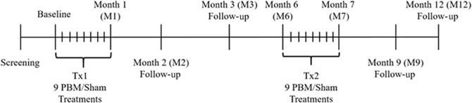
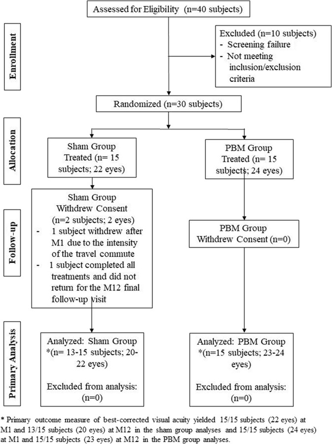
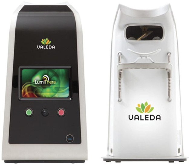
Methods: Thirty subjects (46 eyes) were treated with the Valeda Light Delivery System, wherein subjects underwent two series of treatments (3× per week for 3-4 weeks) over 1 year. Outcome measures included best-corrected visual acuity, contrast sensitivity, microperimetry, central drusen volume and drusen thickness, and quality of life assessments.

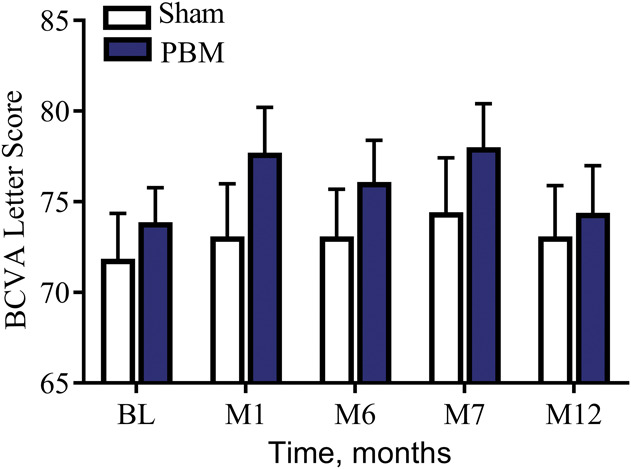
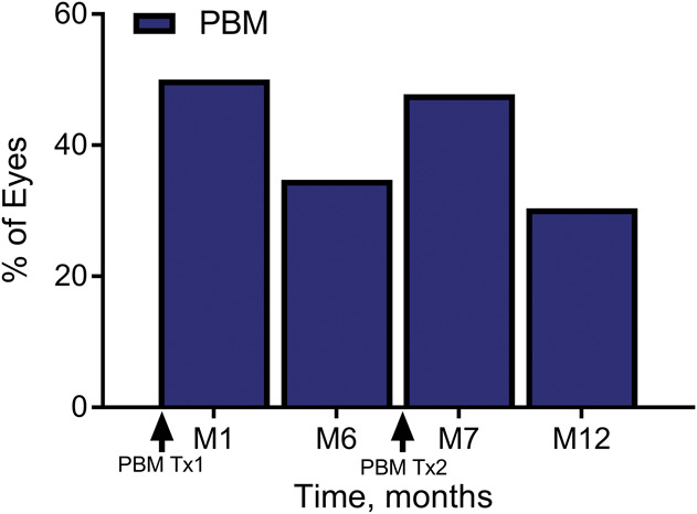
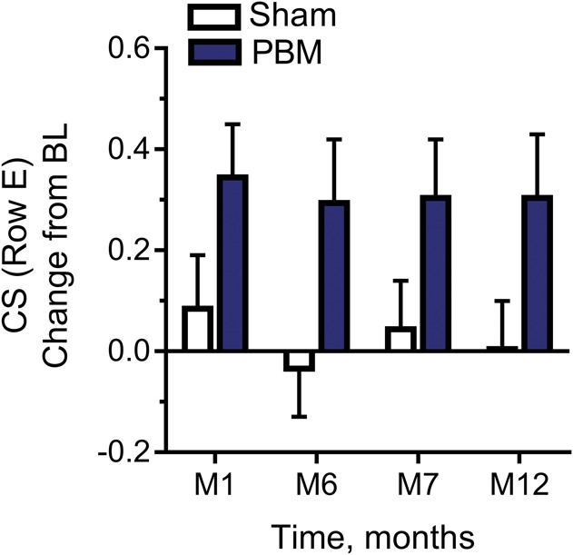
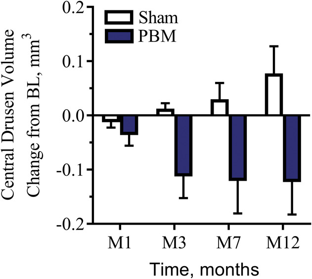
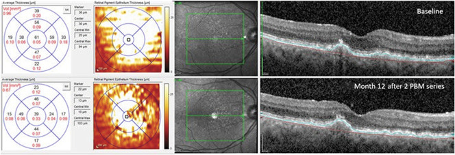
Results: Photobiomodulation-treated subjects showed a best-corrected visual acuity mean letter score gain of 4 letters immediately after each treatment series at Month 1 (M1) and Month 7 (M7). Approximately 50% of PBM-treated subjects showed improvement of ≥5 letters versus 13.6% in sham-treated subjects at M1. High responding subjects (≥5-letter improvement) in the PBM-treated group showed a gain of 8 letters after initial treatment (P < 0.01) and exhibited earlier stages of age-related macular degeneration disease. Statistically significant improvements in contrast sensitivity, central drusen volume, central drusen thickness, and quality of life were observed (P < 0.05). No device-related adverse events were reported.

Conclusion: Photobiomodulation treatment statistically improved clinical and anatomical outcomes with more robust benefits observed in subjects with earlier stages of dry age-related macular degeneration. Repeated PBM treatments are necessary to maintain benefits. These pilot findings support previous reports and suggest the utility of PBM as a safe and effective therapy in subjects with dry age-related macular degeneration.


Conflict of interest statement

R. G. Devenyi and S. N. Markowitz report clinical research agreements and fees from LumiThera Inc, for the conduct of the study. M. R. Munk received lecturer fees from Novartis (Novartis AG) and travel support from Bayer (Bayer AG) and is a consultant for Allergan and Zeiss and reports fees from LumiThera Inc, during the conduct of the study. M. G. Walker and R. Rückert report personal fees and other from LumiThera Inc, during the conduct of the study. LumiThera employees received grant support from National Institutes of Health, National Eye Institute #3R43EY025508 to 01S1. C. E. Tedford and S. E. Stephanie Tedford as well as C. L. Croissant are employees of LumiThera, Inc. The remaining authors have no financial/conflicting interests to disclose.

Figures

Fig. 1.
Fig. 1.
Diagram illustrating the LIGHTSITE I clinical study visit design. Subjects who met the inclusion/exclusion criteria at the screening and baseline visits were enrolled into the study. Subjects received two series of PBM treatments (Tx1 and Tx2) with a total of 9 treatment sessions per series distributed over 3 weeks to 4 weeks. Subjects underwent assessments at subsequent follow-up visits. The study comprised of 24 visits over the course of 1 year.
Fig. 2.
Fig. 2.
LIGHTSITE I consort diagram. Study subject progress through each study phase.
Fig. 3.
Fig. 3.
The Lumithera Valeda Light Delivery System. Illustration of the Valeda front (left) and backside (right).
Fig. 4.
Fig. 4.
The effect of PBM on ETDRS BCVA letter score over-time. As a group, PBM-treated subjects showed an improvement in ∼4 letters immediately after treatment (M1 and M7) which diminished over time demonstrating the need for repeated treatment to maintain clinical benefits.
Fig. 5.
Fig. 5.
The percentage of PBM-treated subjects with a ≥5 letter improvement on ETDRS visual acuity from baseline over time. After each treatment series (M1 and M7), almost one half of all PBM-treated eyes showed ≥5 letter gain (50% of eyes at M1, 46% of eyes at M7). This effect was reduced over time until the next treatment series was initiated. Photobiomodulation-treated eyes that showed ≥5 letter gain were typically in earlier stages of the disease and did not have significant GA with foveola involvement.
Fig. 6.
Fig. 6.
The effect of PBM on contrast sensitivity (Level E, 18.0 CPD) in LogCS change from baseline. Photobiomodulation-treated subjects showed significant improvement in CS at Level E (18 cycles/degree) out to 12 months after treatment, Wilcoxon signed-rank test, P < 0.05.
Fig. 7.
Fig. 7.
The effect of PBM on drusen volume (mm3) over time. Drusen volume increased over time in 100% of the sham-treated subjects. By contrast, 70% of all PBM-treated subjects showed a reduction in drusen. A statistically significant reduction in drusen volume at M12 (LME, P = 0.05) was observed in PBM-treated subjects versus the sham-treated subjects.
Fig. 8.
Fig. 8.
Representative example of anatomical improvement in a PBM-treated eye. This eye was categorized as AREDS 3. Baseline (top) imaging illustrates drusen volume of 0.78 mm3 with a mean central 1-mm drusen thickness of 165 µm. Black numbers indicate the mean thickness of each ETDRS subgrid, and red numbers indicate the corresponding volume (mm3). The maps on the left-hand side depict the color-coded drusen thickness map. Month 12 (bottom) imaging illustrates an overall reduction in volume (0.41 mm3) and mean central thickness (18 µm) after PBM treatment.

References

    1. Wong WL, Su X, Li X, et al. Global prevalence of age-related macular degeneration and disease burden projection for 2020 and 2040: a systematic review and meta-analysis. Lancet Glob Health 2014;2:e106–e116. - PubMed
    1. The Age-Related Eye Disease Study system for classifying age-related macular degeneration from stereoscopic color fundus photographs: the Age-Related Eye Disease Study report number 6. Am J Ophthalmol 2001;132:668–681. - PubMed
    1. Qin S, Rodrigues GA. Progress and perspectives on the role of RPE cell inflammatory responses in the development of age-related macular degeneration. J Inflamm Res 2008;1:49–65. - PMC - PubMed
    1. Chew EY, Chew EY, Clemons TE, et al. Secondary analyses of the effects of lutein/zeaxanthin on age-related macular degeneration progression: AREDS2 report No. 3. JAMA Ophthalmol 2014;132:142–149. - PMC - PubMed
    1. Aronow ME, Chew EY. Age-Related Eye Disease Study 2: perspectives, recommendations, and unanswered questions. Curr Opin Ophthalmol 2014;25:186–190. - PMC - PubMed

Publication types

MeSH terms