. 2019 Aug 12;15(8):e1008002.
doi: 10.1371/journal.ppat.1008002. eCollection 2019 Aug.

Inducible LGALS3BP/90K activates antiviral innate immune responses by targeting TRAF6 and TRAF3 complex

Affiliations

Inducible LGALS3BP/90K activates antiviral innate immune responses by targeting TRAF6 and TRAF3 complex

Gang Xu et al. PLoS Pathog. .

Abstract

The galectin 3 binding protein (LGALS3BP, also known as 90K) is a ubiquitous multifunctional secreted glycoprotein originally identified in cancer progression. It remains unclear how 90K functions in innate immunity during viral infections. In this study, we found that viral infections resulted in elevated levels of 90K. Further studies demonstrated that 90K expression suppressed virus replication by inducing IFN and pro-inflammatory cytokine production. Upon investigating the mechanisms behind this event, we found that 90K functions as a scaffold/adaptor protein to interact with TRAF6, TRAF3, TAK1 and TBK1. Furthermore, 90K enhanced TRAF6 and TRAF3 ubiquitination and served as a specific ubiquitination substrate of TRAF6, leading to transcription factor NF-κB, IRF3 and IRF7 translocation from the cytoplasm to the nucleus. Conclusions: 90K is a virus-induced protein capable of binding with the TRAF6 and TRAF3 complex, leading to IFN and pro-inflammatory production.


Conflict of interest statement

The authors have declared that no competing interests exist.

Figures

Fig 1
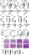
Fig 1. 90K is induced by viruses.
(A-C) C57BL/6 mice MEFs were infected with IAV (MOI = 1) (A), VSV (MOI = 1) (B), HSV-1 (MOI = 1) (C) for indicated times. 90K (left panel) and IFN-α (right panel) expression was quantified by real-time RT-PCR. (D) Experiments were performed as in (A), except that poly (I:C) was used. (E) C57BL/6 mice were infected with IAV (104 TCID50 per mouse, n = 3) for indicated times. 90K mRNA levels in the lung were quantified by real-time RT-PCR. (F) A549 WT cells or IFNAR1-/- cells were infected with IAV (MOI = 1) for indicated times prior to western blot assays. (G) A549 cells were treated with indicated concentrations of IFN-α for 24 hours. The protein level of 90K was analyzed by western blot. All experiments were repeated at least three times with consistent results. In the real-time RT-PCR experiments, the control was designated as 1. Bar graphs present means ± SD, n = 3 (**P < 0.01; *P < 0.05), n.s., not significant.
Fig 2
Fig 2. 90K inhibits virus replication.
(A) A549 cells were transfected with indicated plasmids for 24 hours followed by infection with IAV (MOI = 1) for 24 hours. The relative levels of NP-specific mRNA, cRNA and vRNA were quantified by real-time RT-PCR assay (upper panel) and protein levels of 90K were quantified by western blot (lower panel). (B) Experiments were performed as in (A) except cells were transfected with indicated shRNAs. (C) Huh7 cells were transfected with pHBV-1.3 and an empty vector or HA-90K for 48 hours. The secretion of HBeAg and HBsAg was measured by ELISA (left panel) and the amount of HBV capsid-associated DNA (right panel) was determined by real-time RT-PCR assay. (D) Experiments were performed as in (C) except cells were transfected with indicated shRNAs. (E) A549 cells were transfected with indicated plasmids for 24 hours followed by infection with VSV (MOI = 1) for 24 hours prior to plaque assay (upper panel). The protein levels of 90K were quantified by western blot (lower panel) (F) A549 cells were transfected with indicated shRNAs for 24 hours followed by infection with VSV (MOI = 1) for 8 hours prior to real-time RT-PCR assay (upper panel). The protein levels of 90K were quantified by western blot (lower panel). (G) RD cells were transfected with indicated plasmids for 24 hours followed by infection with EV71 (MOI = 1) for 2 hours prior to real-time RT-PCR assay. (H) Experiments were performed as in (G) except cells were transfected with indicated shRNAs. (I) Hela cells transfected with indicated plasmids for 24 hours followed by infection with HSV-1 for 24 hours prior to real-time RT-PCR assay. (J-M) WT (black square, n = 9) and 90k-/- (red circle, n = 9) mice were intranasally infected with 104 TCID50 of IAV, and body weights were recorded daily (J). Survival curves show data collected until day 14 post-infection (K). The statistical analysis was performed using a log-rank test. Lung/body weight normalized (L) and viral titers (M) in lung tissues were evaluated on indicated times after influenza viral infection. (N) Histological analysis of the lung tissue of WT (n = 3) and 90k-/- mice (n = 3) stained with H&E on days 0, 2, 4, and 6 after intranasal infection with 104 TCID50 IAV. (O) WT and 90k-/- splenocytes were infected with VSV (MOI = 1), ZIKV (MOI = 1), HSV (MOI = 1) or EV71 (MOI = 1) for 12 hours, respectively. The relative levels of nucleocapsid protein (VSV), envelope protein (ZIKV), ICP0 (HSV) or VP1 (EV71) were quantified by real-time RT-PCR assay. All experiments were repeated at least three times with consistent results. In the real-time RT-PCR experiments, the control was designated as 1. Bar graphs present means ± SD, n = 3 (**P < 0.01; *P < 0.05), n.s., not significant.
Fig 3
Fig 3. 90K regulates IAV-induced inflammatory cytokine expression.
(A) A549 cells were transfected with indicated plasmids for 24 hours and infected with IAV (MOI = 1) for indicated times prior to real-time RT-PCR assay. (B-D) qRT-PCR analysis of indicated cytokine mRNA in WT and 90k-/- MEFs (B), splenocytes (C), PBMCs (D) infected with IAV for indicated times. (E) WT (n = 3) and 90k-/- (n = 3) mice were intranasally infected with 104 TCID50 of IAV for indicated times. The relative levels of inflammatory cytokine in mice lung were quantified by real-time RT-PCR assay. All experiments were repeated at least three times with consistent results. In the real-time RT-PCR experiments, the control was designated as 1. Bar graphs present means ± SD, n = 3 (**P < 0.01; *P < 0.05), n.s., not significant.
Fig 4
Fig 4. 90K regulated IRF3, IRF7 and NF-κB activation and ISGs expression.
(A) A549 cells infected with SeV for indicated times prior to western blot analyses. (B) A549 cells transfected with indicated plasmids for 24 hours prior to western blot analyses. (C) A549 cells transfected with indicated plasmids for 24 hours and infected with IAV (MOI = 1) for 6 hours. Cytosolic and nuclear extracts were prepared and subjected to western blot analyses. Lamin A and GAPDH were used as markers for nuclear and cytosolic fractions, respectively. (D-F) WT and 90k-/-MEFs (D), splenocytes (E), PBMCs (F) were infected with IAV for indicated times. The relative levels of ISGs were quantified by real-time RT-PCR or Western blot analyses. (G) WT (n = 3) and 90k-/- (n = 3) mice were intranasally infected with 104 TCID50 of IAV for indicated times. The relative levels of ISGs in mice lung were quantified by real-time RT-PCR. (H and I) A549 cells were transfected with indicated plasmids for 24 hours and infected with IAV (H) or treated with poly (I:C) (I) for indicated times. The relative levels of ISGs were quantified by real-time RT-PCR analyses. (J) A549 cells ere transfected with indicated plasmids for 24 hours and infected with SeV for 24 hours prior to western blot analyses. All experiments were repeated at least three times with consistent results. In the real-time RT-PCR experiments, the control was designated as 1. Bar graphs present means ± SD, n = 3 (**P < 0.01; *P < 0.05), n.s., not significant.
Fig 5
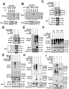
Fig 5. 90K associates with TRAF6 and TAK1.
(A-C) 293T cells were transfected with the indicated plasmids for 48 hours. Coimmunoprecipitation and immunoblots were performed with the indicated antibodies. (D) A549 cells were infected with SeV for the indicated times or left uninfected. Immunoprecipitation and immunoblot analysis were performed with the indicated antibodies. (E-H) Schematic diagram of the full-length and truncated constructs of TAK1, TRAF6 and 90K (upper panel). 293T cells were transfected with the indicated plasmids for 48 hours. (I-K) 293T cells were transfected with the indicated plasmids for 48 hours. Coimmunoprecipitation and immunoblots were performed with the indicated antibodies. All experiments were repeated at least three times with consistent results.
Fig 6
Fig 6. 90K potentiates TRAF6 ubiquitination and is ubiquitinated by TRAF6.
(A and B) 293T cells were transfected with HA or Myc tagged 90K plasmids and Flag-TRAF6 and the indicated ubiquitin plasmids for 48 hours before co-immunoprecipitation and immunoblot analyses were performed with the indicated antibodies. (C) 293T cells transfected with empty vector or Myc-90K for 24 hours. Cells were infected without (left panel) or with (right panel) VSV for 6 hours. Coimmunoprecipitation and immunoblot were performed with the indicated antibodies. (D) 293T cells transfected with empty vector or Myc-90K for 24 hours and infected with VSV for 6 hours. Coimmunoprecipitation and immunoblot were performed with the indicated antibodies. (E and F) 293T cells were transfected with Flag-90K and Myc-Ub or indicated truncated or mutated TRAF6 constructs for 48 hours. Coimmunoprecipitation and immunoblot were performed with the indicated antibodies. (G and H) 293T cells were transfected with the indicated plasmids for 48 hours before co-immunoprecipitation and immunoblot analyses were performed with the indicated antibodies. (I) 293T cells were transfected with Flag-TRAF6 and Myc-Ub or indicated truncated 90K constructs for 48 hours. Coimmunoprecipitation and immunoblot were performed with the indicated antibodies. All experiments were repeated at least three times with consistent results.
Fig 7
Fig 7. 90K interacts with TRAF3 and TBK1, and enhances TRAF3 ubiquitination.
(A and B) 293T cells were transfected with the indicated plasmids for 48 hours. Coimmunoprecipitation and immunoblots were performed with the indicated antibodies. (C and D) 293T cells were transfected with the indicated plasmids for 48 hours. Coimmunoprecipitation and immunoblots were performed with the indicated antibodies. (E-I) 293T cells were transfected with the indicated plasmids and Myc-Ub for 48 hours. Coimmunoprecipitation and immunoblots were performed with the indicated antibodies. All experiments were repeated at least three times with consistent results.
Fig 8
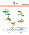
Fig 8. Model of the biological effect of 90K in virus-induced inflammatory cytokine expression.
After virus infection, TRAF6 recruits the newly-expressed 90K through its RING/ZnF domain. Association of the TRAF6 and 90K forms a ubiquitin-conjugating enzyme complex that recruits TAK1. Induced 90K also interacts with TRAF3 and TBK1 to form a complex. Two complexes signal to translocate the transcription factors IRF3/7 and NF-κB, from the cytoplasm to the nucleus for subsequent production of IFN and inflammatory cytokines.

References

    1. Creagh EM, O’Neill LA. TLRs, NLRs and RLRs: a trinity of pathogen sensors that co-operate in innate immunity. Trends Immunol. 2006;27(8):352–7. 10.1016/j.it.2006.06.003 . - DOI - PubMed
    1. Akira S, Takeda K. Toll-like receptor signalling. Nat Rev Immunol. 2004;4(7):499–511. 10.1038/nri1391 . - DOI - PubMed
    1. Cohen P, Strickson S. The role of hybrid ubiquitin chains in the MyD88 and other innate immune signalling pathways. Cell Death Differ. 2017;24(7):1153–9. 10.1038/cdd.2017.17 . - DOI - PMC - PubMed
    1. Ajibade AA, Wang HY, Wang RF. Cell type-specific function of TAK1 in innate immune signaling. Trends Immunol. 2013;34(7):307–16. 10.1016/j.it.2013.03.007 . - DOI - PubMed
    1. Chen ZJ. Ubiquitination in signaling to and activation of IKK. Immunol Rev. 2012;246(1):95–106. 10.1111/j.1600-065X.2012.01108.x . - DOI - PMC - PubMed

Publication types

MeSH terms