. 2019 Jul 25:7:311.
doi: 10.3389/fped.2019.00311. eCollection 2019.

Identification of Mycobacterium tuberculosis Infection in Infants and Children With Partial Discrimination Between Active Disease and Asymptomatic Infection

Affiliations

Identification of Mycobacterium tuberculosis Infection in Infants and Children With Partial Discrimination Between Active Disease and Asymptomatic Infection

Alexandra Dreesman et al. Front Pediatr. .

Abstract

Background: Improved diagnostic tests are needed for the early identification of Mycobacterium tuberculosis-infected young children exposed to an active TB (aTB) index case. We aimed to compare the diagnostic accuracy of new blood-based tests to that of the tuberculin skin test (TST) for the identification of all infected children and for a potential differentiation between aTB and latent TB infection (LTBI). Methods: 144 children exposed to a patient with aTB were included, and those who met all inclusion criteria (130/144) were classified in three groups based on results from classical investigations: non-infected (NI: n = 69, 53%, median age 10 months), LTBI (n = 28, 22%, median age 96 months), aTB disease (n = 33, 25%, median age 24 months). The first whole blood assay consisted of a 7-days in vitro stimulation of blood with four different mycobacterial antigens (40 μl/condition), followed by flow cytometric measurement of the proportions of blast cells appearing among lymphocytes as a result of their specific activation. Thresholds of positivity were determined by Receiver Operating Characteristic (ROC) curve analysis (results of NI children vs. children with LTBI/aTB) in order to identify infected children in a first stage. Other cut-offs were determined to discriminate subgroups of infected children in a second step (results from children with aTB/LTBI). Analysis of blood monocytes and dendritic cell subsets was performed on 100 μl of blood for 25 of these children as a second test in a pilot study. Results: Combining the results of the blast-induced CD3+ T lymphocytes by Heparin-Binding Haemagglutinin and by Culture Filtrate Protein-10 identified all but one infected children (sensitivity 98.2% and specificity 86.9%, compared to 93.4 and 100% for the TST). Further identification among infected children of those with aTB was best achieved by the results of blast-induced CD8+ T lymphocytes by purified protein derivative (sensitivity for localized aTB: 61.9%, specificity 96.3%), whereas high proportions of blood type 2 myeloid dendritic cells (mDC) were a hallmark of LTBI. Conclusions: New blood-based tests requiring a very small volume allow the accurate identification of M. tuberculosis-infected young children among exposed children and are promising to guide the clinical classification of children with aTB or LTBI.

Keywords: FASCIA; Mycobacterium tuberculosis; active tuberculosis; children; dendritic cells; diagnosis; latent infection; lymphoblasts.


Figures
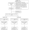
Figure 1
Figure 1
STARD diagram reporting the flow of children for the evaluation of the combined HBHA- and CFP-10-induced CD3+ blasts lymphocytes for the identification of M. tuberculosis-infected children (LTBI/aTB). The aim of this index test was to correctly classify children exposed to an index case of aTB disease in two groups: non-infected children, and infected children comprising both children with LTBI and those with aTB disease. The reference standard test was the tuberculin skin test (TST) performed at the inclusion of the child in the study (first TST), the TST being the reference test recommended in Belgium and in most European countries at least for children younger than 5 years.
Figure 2
Figure 2
Percentages of CD3+ blasts induced by mycobacterial antigens in M. tuberculosis-infected and in non-infected children. Ten-fold diluted whole blood was incubated during 7 days at 37°C with PPD (4 μg/ml), HBHA (10 μg/ml), ESAT-6 (10 μg/ml), or CFP-10 (5 μg/ml), as indicated, before labeling the cells with anti-CD3 monoclonal antibody and analysis of the percentages of CD3+ blasts by flow cytometry. Each symbol represents the value from an individual subject after deduction of the percentage obtained in non-stimulated condition, and the horizontal lines indicate the medians. The dotted horizontal lines indicate the cut-off values determined by ROC curve analysis. Values obtained for non-infected (NI) children were compared to those from infected children (LTBI/aTB) by Mann-Whitney U test.
Figure 3
Figure 3
Combined analysis of the percentages of HBHA- and CFP-10-induced CD3+ blasts in M. tuberculosis-infected children (LTBI/aTB). Ten-fold diluted whole blood was incubated during 7 days at 37°C with HBHA (10 μg/ml) or CFP-10 (5 μg/ml), as indicated, before labeling the cells with anti-CD3 monoclonal antibody and analysis of the percentages of CD3+ blasts by flow cytometry. (A) Results from M. tuberculosis-infected children (LTBI and aTB combined), and (B) results from non-infected children, each symbol representing the value obtained for an individual subject after deduction of the percentage obtained in the non-stimulated condition. The dotted lines indicate the cut-offs chosen for the two tests.
Figure 4
Figure 4
Comparison of the percentages of T cell blasts induced by mycobacterial antigens between children with LTBI and those with aTB. Ten-fold diluted whole blood was incubated during 7 days at 37°C with PPD (4 μg/ml), HBHA (10 μg/ml), ESAT-6 (10 μg/ml) or CFP-10 (5 μg/ml), as indicated, and the cells were labeled with anti-CD3, anti-CD4, and anti-CD8 monoclonal antibodies. The percentages of CD3+ (upper panels), CD4+ (middle panels) and CD8+ (lower panels) blasts were determined by flow cytometry. Each symbol represents the value from an individual child after deduction of the percentage obtained in the non-stimulated condition. The horizontal filled lines indicate the medians. For the tests providing statistically significant differences between the two groups of infected children, horizontal dotted lines were added to indicate the cut-off of positivity. Statistical analysis was performed by Mann-Whitney U test. LTBI, latently TB infected; aTB, active tuberculosis.
Figure 5
Figure 5
STARD diagram reporting the flow of M. tuberculosis-infected children (LTBI/aTB) for the evaluation of the PPD-induced CD8+ blasts lymphocytes for the identification of children with aTB on the one hand and those with LTBI on the other hand. The reference standard test was the tuberculin skin test (TST) performed in a first diagnostic stage at the time of inclusion in the study. This diagram reports results obtained for the 56 M. tuberculosis-infected children (LTBI/aTB) detected by the combined HBHA- and CFP-10-induced CD3+ FASCIA as reported in Figure 1.
Figure 6
Figure 6
Proportions of circulating plasmacytoïd and myeloïd DC subsets in M. tuberculosis-infected compared to non-infected children. (A,B) The proportions of pDC and of total mDC were evaluated in the peripheral blood of non-infected children (NI), children with a latent TB infection (LTBI), or with active TB (aTB) and the results were expressed as percentages among CD45+ cells (upper panels). (C,D) The proportions of type 1 and type 2 mDC subsets are those among the total mDC (lower panels). Horizontal lines represent the medians of percentages. Statistical analysis was performed by a Kruskal-Wallis test followed by Dunn's tests.

References

    1. WHO Global Tuberculosis Report. WHO (2018). Available online at: http://www.who.int/tb/publications/global_report/en/.
    1. Jenkins HE. Global burden of childhood tuberculosis. Pneumonia. (2016) 8:24. 10.1186/s41479-016-0018-6 - DOI - PMC - PubMed
    1. Perez-Velez CM, Marais BJ. Tuberculosis in children. N Engl J Med. (2012) 367:348–61. 10.1056/NEJMra1008049 - DOI - PubMed
    1. Vanden Driessche K, Persson A, Marais BJ, Fink PJ, Urdahl KB. Immune vulnerability of infants to tuberculosis. Clin Dev Immunol. (2013) 2013:781320. 10.1155/2013/781320 - DOI - PMC - PubMed
    1. O'Garra A, Redford PS, McNab FW, Bloom CI, Wilkinson RJ, Berry MPR. The immune response in tuberculosis. Annu Rev Immunol. (2013) 31:475–527. 10.1146/annurev-immunol-032712-095939 - DOI - PubMed