Sonographic Detection of a Torsed Meckel's Diverticulum Misinterpreted as Acute Appendicitis
- PMID: 31404174
- PMCID: PMC6682254
- DOI: 10.5811/cpcem.2019.5.42976
Sonographic Detection of a Torsed Meckel's Diverticulum Misinterpreted as Acute Appendicitis
Abstract
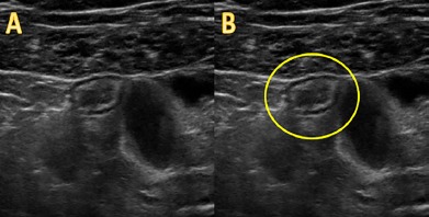
A 38-year-old female presented to the emergency department (ED) with acute-onset right lower quadrant abdominal pain following two days of nausea and vomiting. Physical examination revealed right lower quadrant tenderness to palpation, rebound tenderness, and guarding. Point-of-care ultrasound (POCUS) of the right lower abdomen was performed and interpreted as probable appendicitis. However, upon laparoscopic examination of the abdomen, a benign-appearing appendix was visualized. Further investigation revealed the source of the patient's pain to be a torsed Meckel's diverticulum. Although rare, a torsed and inflamed Meckel's diverticulum can be visualized by POCUS in the ED without the need for further imaging or delay.
Conflict of interest statement
Conflicts of Interest: By the CPC-EM article submission agreement, all authors are required to disclose all affiliations, funding sources and financial or management relationships that could be perceived as potential sources of bias. The authors disclosed none.
Figures