Review
. 2019 Jul;8(Suppl 1):S22-S27.
doi: 10.21037/gs.2019.03.01.

Fluorescence techniques in adrenal surgery

Affiliations
Review

Fluorescence techniques in adrenal surgery

Edwina C Moore et al. Gland Surg. 2019 Jul.

Abstract

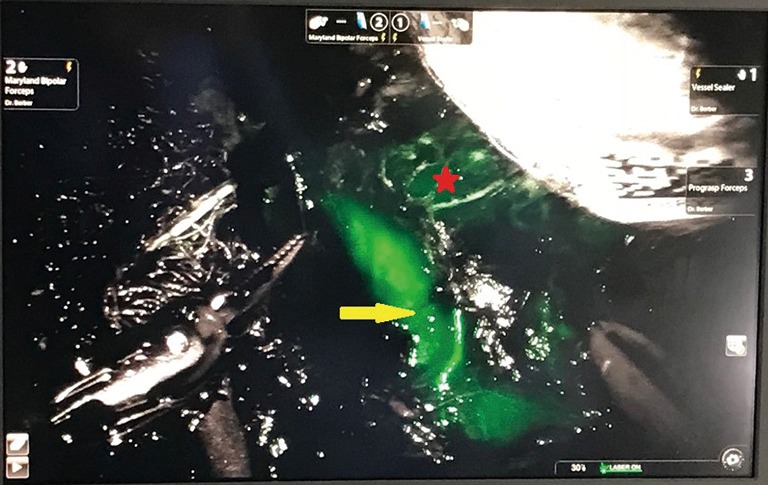
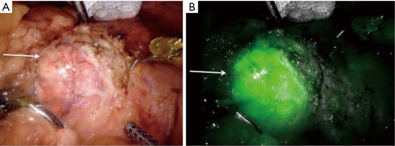
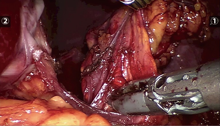
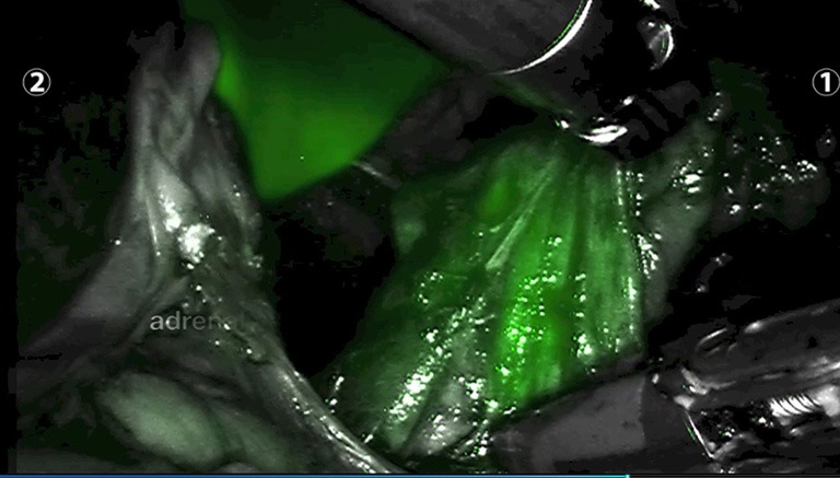
This chapter describes the use of fluorescence via indocyanine green (ICG) in minimally invasive adrenal surgery (laparoscopic and robotic). ICG is a non-toxic dye that can aid identification of vascular structures and parenchymal tissue planes in real time. The primary utility of ICG fluorescence in adrenal surgery is to help delineate the margins of resection, to guide a more precise operation. In particular, for patients with bilateral adrenal disease or a heredity associated with high risk of recurrence (e.g., VHL, MEN2a) this may facilitate subtotal adrenal resection (e.g., cortical sparing adrenalectomy), obviating the incidence of iatrogenic adrenal insufficiency and its numerous sequelae including lifelong hormone supplementation, osteoporosis and risk of Addisonian crisis.

Keywords: Adrenal surgery; fluorescence angiography; indocyanine green (ICG); laparoscopy; near infrared imaging (NIR imaging).


Conflict of interest statement

Conflicts of Interest: The authors have no conflicts of interest to declare.

Figures

Figure 1
Figure 1
Photograph from robotic platform demonstrating a plane of dissection between the left renal vein (yellow arrow) and left adrenal tumour (red star) following ICG administration (original image).
Figure 2
Figure 2
Photograph demonstrating the utility of ICG in delineating adrenal cortical tumours (yellow arrow) from the surrounding retroperitoneum, before ICG administration (A) and after ICG administration (B) (original image). ICG, indocyanine green.
Figure 3
Figure 3
Photograph demonstrating hyper-fluorescence of adrenal cortical tumours (white arrow) before (A) and after ICG administration (B) (original image). ICG, indocyanine green.
Figure 4
Figure 4
Photograph of ICG vial and sterile (distilled) water required for constitution prior to intravenous administration (original image). ICG, indocyanine green.
Figure 5
Figure 5
Photograph prior to ICG administration of left adrenal gland (original image). ICG, indocyanine green.
Figure 6
Figure 6
Photograph after ICG administration of left adrenal gland and hyper-fluorescent liver parenchyma in the background) (original image). ICG, indocyanine green.

References

    1. Yannuzzi LA, Slakter JS, Sorenson JA, et al. Digital indocyanine green videoangiography and choroidal neovascularization. Retina 1992;12:191-223. 10.1097/00006982-199212030-00003 - DOI - PubMed
    1. Ishizawa T, Bandai Y, Kokudo N. Fluorescent cholangiography using indocyanine green for laparoscopic cholecystectomy: an initial experience. Arch Surg 2009;144:381-2. 10.1001/archsurg.2009.9 - DOI - PubMed
    1. Ishizawa T, Bandai Y, Ijichi M, et al. Fluorescent cholangiography illuminating the biliary tree during laparoscopic cholecystectomy. Br J Surg 2010;97:1369-77. 10.1002/bjs.7125 - DOI - PubMed
    1. Tobis S, Knopf JK, Silvers CR, et al. Near infra-red fluorescence imaging after indocyanine green: initial clinical experience with open partial nephrectomy for renal cortical tumours. Urology 2012;79:958-64. 10.1016/j.urology.2011.10.016 - DOI - PubMed
    1. Kahramangil B, Berber E. The use of near infra-red fluorescence imaging in endocrine surgical procedures. J Surg Oncol 2017;115:848-55. 10.1002/jso.24583 - DOI - PubMed