. 2019 Jul 25:10:804.
doi: 10.3389/fneur.2019.00804. eCollection 2019.

Cerebrospinal Fluid Findings in Patients With Autoimmune Encephalitis-A Systematic Analysis

Affiliations

Cerebrospinal Fluid Findings in Patients With Autoimmune Encephalitis-A Systematic Analysis

Tetyana Blinder et al. Front Neurol. .

Abstract

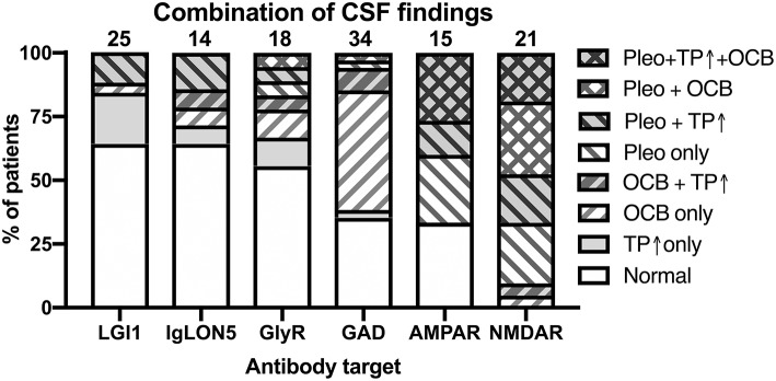
Autoimmune encephalitides (AIE) comprise a group of inflammatory diseases of the central nervous system (CNS), which can be further characterized by the presence of different antineuronal antibodies. Recently, a clinical approach for diagnostic criteria for the suspected diagnosis of AIE as well as definitive AIE were proposed. These are intended to guide physicians when to order the antineuronal antibody testing and/or facilitate early diagnosis even prior to the availability of the specific disease-confirming test results to facilitate prompt treatment. These diagnostic criteria also include the results of basic cerebrospinal fluid (CSF) analysis. However, the different antibody-defined AIE subtypes might be highly distinct with regard to their immune pathophysiology, e.g., the pre-dominance of specific IgG subclasses, IgG1, or IgG4, or frequency of paraneoplastic compared to idiopathic origin. Thus, it is conceivable that the results of basic CSF analysis might also be very different. However, this has not been explored systematically. Here, we systematically reviewed the literature about the 10 most important AIE subtypes, AIE with antibodies against NMDA, AMPA, glycine, GABAA, and GABAB receptors as well as DPPX, CASPR2, LGI1, IgLON5, or glutamate decarboxylase (GAD), with respect to the reported basic CSF findings comprising CSF leukocyte count, total protein, and the presence of oligoclonal bands (OCB) restricted to the CSF as a sensitive measure for intrathecal IgG synthesis. Our results indicate that these basic CSF findings are profoundly different among the 10 different AIE subtypes. Whereas, AIEs with antibodies against NMDA, GABAB, and AMPA receptors as well as DPPX show rather frequent inflammatory CSF changes, in AIEs with either CASPR2, LGI1, GABAA, or glycine receptor antibodies CSF findings were mostly normal. Two subtypes, AIEs defined by either GAD, or IgLON5 antibodies, did not fit into this general pattern. In AIE with GAD antibodies, positive OCBs in the absence of other changes were typical, while the CSF in IgLON5 antibody-positive AIE was characterized by elevated protein.

Keywords: GAD antibodies; LGI1 antibodies; NMDAR antibodies; antineuronal antibodies; autoimmune encephalitis; cerebrospinal fluid; oligoclonal bands; pleocytosis.


Figures

Figure 1
Figure 1
Study profile.
Figure 2
Figure 2
Demographic characteristics of patients with autoimmune encephalitis subtypes defined by 10 different antibodies. (A) Distribution of individual reported ages in patients with autoimmune encephalitides (AIE) of the antibody-defined subtypes. The distribution is depicted as violin plots. Patients younger than 12 years were excluded. The median age is indicated as bold line, the interquartile range is indicated by fine lines. Note the bimodal age distribution of the patients with GABAAR antibodies. (B) Gender distribution of the combined groups of patients with group wise data about the gender distribution available. The percentage of females is depicted as the black part of the bar.
Figure 3
Figure 3
Differential frequency of CSF pleocytosis and CSF cell count in patients with different antibody-defined autoimmune encephalitis subtypes. (A) The cumulative percentage of the patient groups identified in the literature with CSF pleocytosis. The percentage of patients with reported CSF pleocytosis is indicted as black. The total number of patients is indicated above each bar. (B) Distribution of reported CSF cell counts in individual patients with antibody-defined autoimmune encephalitis (AIE) subtypes and pleocytosis. The median cell count identified as a bold line. The origin of the y-axis is set to 5 cells/μl, the lowest pathological cell count.
Figure 4
Figure 4
Reported frequencies of increased protein and oligoclonal IgG in the CSF of patients with different antibody-defined autoimmune encephalitis subtypes. (A) The cumulative percentage of the antibody-defined AIE patient groups identified in the literature with increased CSF protein. The percentage of patients with reported CSF protein is indicated as black. The total number of patients is indicated above each bar. (B) Distribution of reported pathological CSF protein values in individual patients with antibody-defined autoimmune encephalitis (AIE) subtypes. The median cell count identified by a bold line. The origin of the y-axis is set to 450 mg/l as upper normal limit for CSF protein. (C) The cumulative percentage of the antibody-defined AIE patient groups identified in the literature with isolated oligoclonal bands (OCB) in the CSF. The percentage of patients with positive OCB is indicated as black. The total number of patients is indicated above each bar.
Figure 5
Figure 5
Relationships of the reported frequencies of pleocytosis, elevated protein, and presence of oligoclonal IgG in the CSF of patients with different antibody-defined autoimmune encephalitis subtypes. Graphs depict the cumulative frequencies of reported increased CSF cell counts plotted against the frequencies of reportedly positive oligoclonal IgG [OCB positive, (A)], increased CSF protein against pleocytosis (B) as well as the frequency of increased protein plotted against OCB positivity (C). The antibody-defined autoimmune encephalitis (AIE) subtypes were grouped as those with infrequent pathological changes (empty symbol: LGI1, GABAAR, GlyR, CASPR2), frequent pathological changes (black symbols: AMPAR, DPPX, GABABR, NMDAR) as well as those rather distinct patterns of CSF pathologies (gray symbols: IgLON5, GAD). Compared to the rare occurrence of CSF pleocytosis and positive OCB, CSF of patients with IgLON5 antibodies were frequently reported to exhibit elevated CSF protein. CSF findings in patients with GAD antibodies are characterized by a high frequency of positive OCB while pleocytosis and elevated CSF protein rarely occur.
Figure 6
Figure 6
Combination of pathological CSF findings in patients with antibody-defined autoimmune encephalitis. Patients with individual data with regard to all three basic CSF analyses–presence of pleocytosis, elevated CSF protein and positive OCBs–were analyzed for the frequencies of the eight possible combinations of all the tree pathologies. The order of the different antibodies was determined by the percentage of patients with normal or not definitely inflammatory CSF findings (increased protein only). Pleo, pleocytosis; TP ↑, total protein increased; OCB, positive isolated oligoclonal bands in CSF. Diagonal stripes upwards from left to right: positive OCB, downwards: pleocytosis.

References

    1. Dalmau J, Graus F. Antibody-mediated encephalitis. N Engl J Med. (2018) 378:840–51. 10.1056/NEJMra1708712 - DOI - PubMed
    1. Titulaer MJ, McCracken L, Gabilondo I, Armangue T, Glaser C, Iizuka T, et al. . Treatment and prognostic factors for long-term outcome in patients with anti-NMDA receptor encephalitis: an observational cohort study. Lancet Neurol. (2013) 12:157–65. 10.1016/S1474-4422(12)70310-1 - DOI - PMC - PubMed
    1. Graus F, Titulaer MJ, Balu R, Benseler S, Bien CG, Cellucci T, et al. . A clinical approach to diagnosis of autoimmune encephalitis. Lancet Neurol. (2016) 15:391–404. 10.1016/S1474-4422(15)00401-9 - DOI - PMC - PubMed
    1. Dalmau J, Gleichman AJ, Hughes EG, Rossi JE, Peng X, Lai M, et al. . Anti-NMDA-receptor encephalitis: case series and analysis of the effects of antibodies. Lancet Neurol. (2008) 7:1091–8. 10.1016/S1474-4422(08)70224-2 - DOI - PMC - PubMed
    1. Lai M, Huijbers MG, Lancaster E, Graus F, Bataller L, Balice-Gordon R, et al. . Investigation of LGI1 as the antigen in limbic encephalitis previously attributed to potassium channels: a case series. Lancet Neurol. (2010) 9:776–85. 10.1016/S1474-4422(10)70137-X - DOI - PMC - PubMed

Publication types

LinkOut - more resources