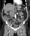
An Unusual Presentation of Cholecystoduodenal Fistula: Abdominal Pain out of Proportion to Exam
- PMID: 31404365
- PMCID: PMC6682232
- DOI: 10.5811/cpcem.2019.4.42686
An Unusual Presentation of Cholecystoduodenal Fistula: Abdominal Pain out of Proportion to Exam
Abstract
Cholecystoduodenal fistula (CDF) is a rare complication of gallbladder disease. Clinical presentation is variable, and preoperative diagnosis is challenging due to the non-specific symptoms of CDF. We discuss a 61-year-old male with a history of atrial fibrillation who presented with severe abdominal pain out of proportion to exam. The patient was diagnosed promptly and successfully managed non-operatively. This case presentation emphasizes the need to maintain a broad differential diagnosis for abdominal pain out of proportion to exam, with the possibility of a biliary-enteric fistula as a possible cause. It also stresses the importance of a multimodality imaging approach to arrive at a final diagnosis.
Conflict of interest statement
Conflicts of Interest: By the CPC-EM article submission agreement, all authors are required to disclose all affiliations, funding sources and financial or management relationships that could be perceived as potential sources of bias. The authors disclosed none.
Figures


References
-
- Glenn F, Reed C, Grafe WR. Biliary enteric fistula. Surg Gynecol Obstet. 1981;153(4):527–31. - PubMed
-
- Chowbey PK, Bandyopadhyay SK, Sharma A, et al. Laparoscopic management of cholecystoenteric fistulas. J Laparoendosc Adv Surg Tech A. 2006;16(5):467–72. - PubMed
-
- Sharma A, Sullivan M, English H, et al. Laparoscopic repair of cholecystoduodenal fistulae. Surg Laparosc Endosc. 1994;4(6):433–5. - PubMed
-
- Abou-Saif A, Al-Kawas FH. Complications of gallstone disease: Mirizzi syndrome, cholecystocholedochal fistula, and gallstone ileus. Am J Gastroenterol. 2002;97(2):249–54. - PubMed
