

Utilizing fourth-generation endocytoscopy and the 'enlarged nuclear sign' for in vivo diagnosis of early gastric cancer
- PMID: 31404432
- PMCID: PMC6687508
- DOI: 10.1055/a-0957-2866
Utilizing fourth-generation endocytoscopy and the 'enlarged nuclear sign' for in vivo diagnosis of early gastric cancer
Abstract
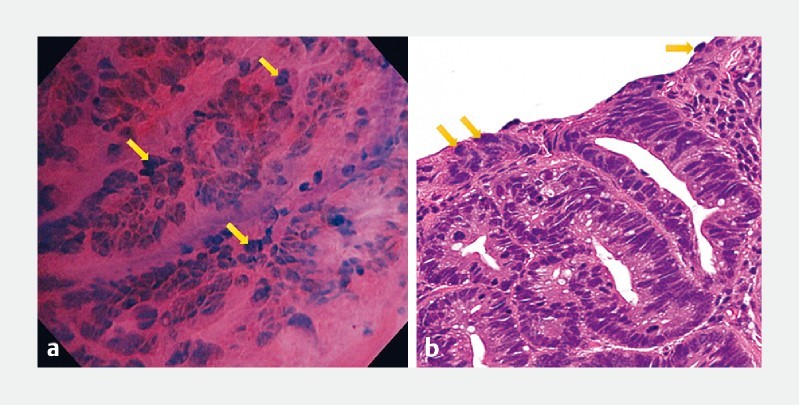
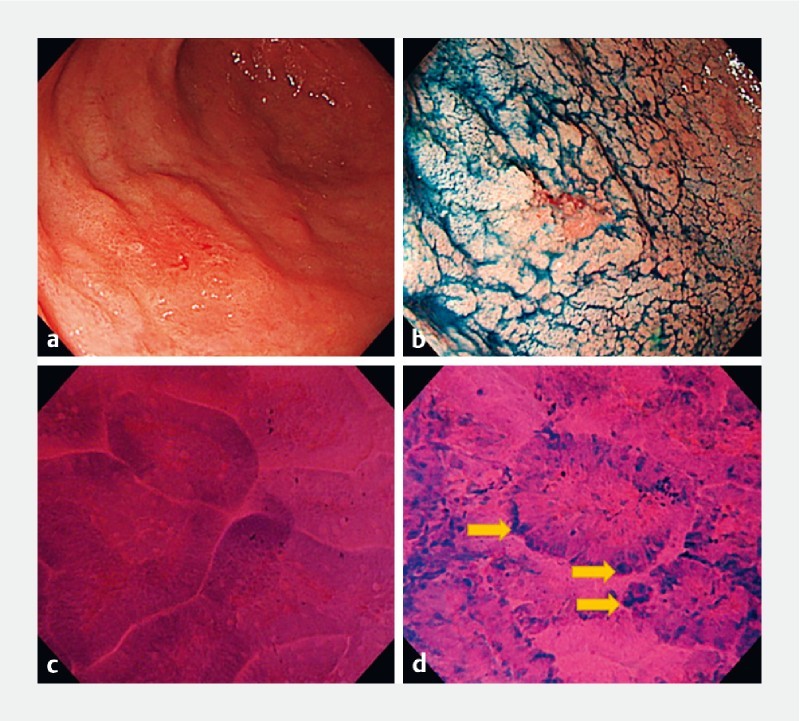
Background and study aims Fourth-generation endocytoscopy is an ultra-high magnification endoscopic technique designed to provide excellent quality in vivo histologic assessment of gastrointestinal lesions. This study aims to evaluate the diagnostic accuracy of endocytoscopy in early gastric cancer diagnosis. Patients and methods A single-center, retrospective analysis of prospectively collected data from all gastric endocytoscopic examinations was conducted. Two expert endoscopists, blinded to white-light and narrow-band imaging findings as well as histopathologic diagnosis, independently reviewed and diagnosed all endocytoscopic images. A newly recognized "enlarged nuclear sign" was detected, and its implication in early gastric cancer diagnosis was evaluated. The diagnostic performance of fourth-generation endocytoscopy was assessed while using the gold standard histopathology as a reference. Results Forty-three patients (mean age±SD, 72.6 ± 12.1 years; 31 males) were enrolled. Based on histopathology, 23 had well-differentiated adenocarcinomas, four adenomas, and 16 non-neoplastic lesions. The sensitivity, specificity, and accuracy of fourth-generation endocytoscopy for gastric cancer diagnosis were 87.0 % (95 % CI: 67.9 - 95.5), 80.0 % (95 % CI: 58.4 - 91.9), and 83.7 % (95 % CI: 70.0 - 91.9) by endoscopist A; and 91.3 % (95 % CI: 73.2 - 97.6), 75.0 % (95 % CI: 53.1 - 88.8), and 83.7 % (95 % CI: 70.0 - 91.9) by endoscopist B. The inter-observer agreement, Kappa statistic = 0.71 (95 % CI: 0.50 - 0.93), was good. The sensitivity, specificity, and accuracy of the enlarged nuclear sign for early gastric cancer diagnosis were 87.0 % (95 % CI: 67.9 - 95.5), 95.0 % (95 % CI: 76.4 - 99.1), and 90.7 % (95 % CI: 78.4 - 96.3) by endoscopist A; and 82.6 % (95 % CI: 62.9 - 93.0), 85.0 % (95 % CI: 64.0 - 94.8), and 83.7 % (95 % CI: 70.0 - 91.9) by endoscopist B. The inter-observer agreement, Kappa statistic = 0.68 (95 % CI: 0.51 - 0.89) was good. Conclusion: Fourth-generation endocytoscopy appears to aid in the diagnosis of early gastric cancer, particularly well-differentiated adenocarcinomas, due to its good diagnostic accuracy and identification of the "enlarged nuclear sign," and deserves further evaluation in future studies.
Conflict of interest statement
Dr. Manolakis is a Hellenic Society of Gastroenterology grant holder. Dr. Rodriguez de Santiago is a Ramón y Cajal Health Research Institute grant holder.
Figures
References
-
- Bray F, Ferlay J, Soerjomataram I et al. Global cancer statistics 2018: GLOBOCAN estimates of incidence and mortality worldwide for 36 cancers in 185 countries. Ca Cancer J Clin. 2018;0:1–31. - PubMed
-
- Kaise M, Ohkura Y, Iizuka T et al. Endocytoscopy is a promising modality with high diagnostic accuracy for gastric cancer. Endoscopy. 2015;47:19–25. - PubMed
-
- Kumagai Y, Kawada K, Takubo K et al. Ultra-high magnification endoscopy (endocytoscopy system) for examination of esophageal lesions. Gastroenterol Endosc. 2017;59:209–218.
-
- Kumagai Y, Takubo K, Kawada K et al. A newly developed continuous zoom-focus endocytoscope. Endoscopy. 2017;49:176–180. - PubMed
LinkOut - more resources
Full Text Sources