

Automated Segmentation of Tissues Using CT and MRI: A Systematic Review
- PMID: 31405724
- PMCID: PMC6878163
- DOI: 10.1016/j.acra.2019.07.006
Automated Segmentation of Tissues Using CT and MRI: A Systematic Review
Abstract
Rationale and objectives: The automated segmentation of organs and tissues throughout the body using computed tomography and magnetic resonance imaging has been rapidly increasing. Research into many medical conditions has benefited greatly from these approaches by allowing the development of more rapid and reproducible quantitative imaging markers. These markers have been used to help diagnose disease, determine prognosis, select patients for therapy, and follow responses to therapy. Because some of these tools are now transitioning from research environments to clinical practice, it is important for radiologists to become familiar with various methods used for automated segmentation.
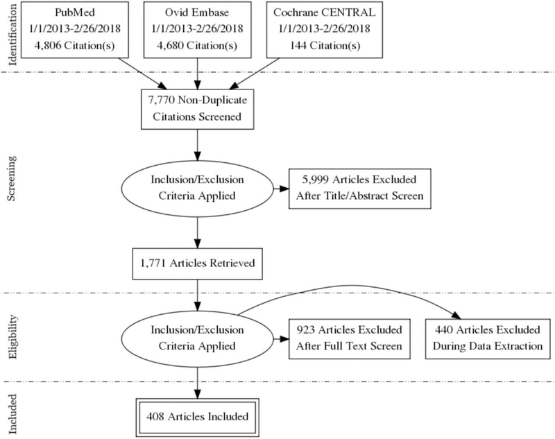
Materials and methods: The Radiology Research Alliance of the Association of University Radiologists convened an Automated Segmentation Task Force to conduct a systematic review of the peer-reviewed literature on this topic.
Results: The systematic review presented here includes 408 studies and discusses various approaches to automated segmentation using computed tomography and magnetic resonance imaging for neurologic, thoracic, abdominal, musculoskeletal, and breast imaging applications.
Conclusion: These insights should help prepare radiologists to better evaluate automated segmentation tools and apply them not only to research, but eventually to clinical practice.
Keywords: CT; MRI; Machine learning; Quantitative imaging; Segmentation.
Copyright © 2019 The Association of University Radiologists. Published by Elsevier Inc. All rights reserved.
Figures
References
Appendix B: Manuscripts Included in Systematic Review
B1. Neurosegmentation
-
- Abbasi S, Tajeripour F. Detection of brain tumor in 3D MRI images using local binary patterns and histogram orientation gradient. Neurocomputing. 2017; 219:526–535.
-
- Adamson C, Beare R, Walterfang M, Seal M. Software Pipeline for Midsagittal Corpus Callosum Thickness Profile Processing: Automated Segmentation, Manual Editor, Thickness Profile Generator, Group-Wise Statistical Comparison and Results Display. Neuroinformatics. 2014; 12(4):595–614. - PubMed
B2. Thoracic Segmentation
-
- Abbas Q Segmentation of differential structures on computed tomography images for diagnosis lung-related diseases. Biomed Signal Process Control. 2017;33:325–34.
-
- Albà X, Figueras I Ventura RM, Lekadir K, Tobon-Gomez C, Hoogendoorn C, Frangi AF. Automatic cardiac LV Segmentation in MRI using modified graph cuts with smoothness and interslice constraints. Magn Reson Med. 2014;72(6):1775–84. - PubMed
-
- Albà X, Lekadir K, Pereañez M, Medrano-Gracia P, Young AA, Frangi AF. Automatic initialization and quality control of large-scale cardiac MRI segmentations. Med Image Anal. 2018;43:129–41. - PubMed
-
- Anders K, Achenbach S, Petit I, Daniel WG, Uder M, Pflederer T. Accuracy of automated software-guided detection of significant coronary artery stenosis by CT angiography: Comparison with invasive catheterisation. Eur Radiol. 2013;23(5):1218–25. - PubMed
-
- Anthimopoulos M, Christodoulidis S, Ebner L, Christe A, Mougiakakou S. Lung Pattern Classification for Interstitial Lung Diseases Using a Deep Convolutional Neural Network. IEEE Trans Med Imaging. 2016;35(5):1207–16. - PubMed
B3. Abdominal Segmentation
-
- Acosta O, Mylona E, Le Dain M, Voisin C, Lizee T, Rigaud B, et al. Multi-atlas-based segmentation of prostatic urethra from planning CT imaging to quantify dose distribution in prostate cancer radiotherapy. Radiother Oncol. 2017;125(3):492–9. - PubMed
-
- Anter AMH AE; ElSoud MA; Azar AT Automatic liver parenchyma segmentation system from abdominal CT scans using hybrid techniques. International Journal of Biomedical Engineering and Technology.01 January 2015;17(2):148–167.
B4. Musculoskeletal Segmentation
-
- Almeida DF, Ruben RB, Folgado J, Fernandes PR, Audenaert E, Verhegghe B, et al. Fully automatic segmentation of femurs with medullary canal definition in high and in low resolution CT scans. Med Eng Phys. 2016;38(12):1474–80. - PubMed
-
- Anas EM, Rasoulian A, Seitel A, Darras K, Wilson D, John PS, et al. Automatic Segmentation of Wrist Bones in CT Using a Statistical Wrist Shape + Pose Model. IEEE Trans Med Imaging. 2016;35(8):1789–801. - PubMed
-
- Athertya JS, Saravana Kumar G. Automatic segmentation of vertebral contours from CT images using fuzzy corners. Comput Biol Med. 2016;72:75–89. - PubMed
B5. Breast Segmentation
-
- DalmƖş MU, Litjens G, Holland K, Setio A, Mann R, Karssemeijer N, Gubern Mérida A. Using deep learning to segment breast and fibroglandular tissue in MRI volumes. Medical physics. 2017. February 1;44(2):533–46. - PubMed
B6. Adipose Tissue Segmentation
-
- Addeman BT, Kutty S, Perkins TG, Soliman AS, Wiens CN, McCurdy CM, Beaton MD, Hegele RA, McKenzie CA. Validation of volumetric and single slice MRI adipose analysis using a novel fully automated segmentation method. Journal of Magnetic Resonance Imaging. 2015. January;41(1):233–41. - PubMed
-
- Decazes P, Rouquette A, Chetrit A, Vera P, Gardin I. Automatic measurement of the total visceral adipose tissue from computed tomography images by using a multi-atlas segmentation method. Journal of computer assisted tomography. 2018. January 1;42(1):139–45. - PubMed
-
- Ding X, Terzopoulos D, Diaz Zamudio M, Berman DS, Slomka PJ, Dey D. Automated pericardium delineation and epicardial fat volume quantification from noncontrast CT. Medical physics. 2015. September 1;42(9):5015–26. - PubMed
-
- Fallah F, Machann J, Martirosian P, Bamberg F, Schick F, Yang B. Comparison of T1-weighted 2D TSE, 3D SPGR, and two-point 3D Dixon MRI for automated segmentation of visceral adipose tissue at 3 Tesla. Magnetic Resonance Materials in Physics, Biology and Medicine. 2017. April 1;30(2):139–51. - PubMed
References
-
- McBee MP, Awan OA, Colucci AT, Ghobadi CW, Kadom N, Kansagra AP, Tridandapani S, Auffermann WF. Deep learning in radiology. Academic Radiology. 2018. November 1;25(11):1472–80. - PubMed
-
- Lundervold AS, Lundervold A. An overview of deep learning in medical imaging focusing on MRI. Zeitschrift für Medizinische Physik. 2018. December 13. - PubMed
-
- Stanzione A, Cuocolo R, Cocozza S, Romeo V, Persico F, Fusco F, Longo N, Brunetti A, Imbriaco M. Detection of Extraprostatic Extension of Cancer on Biparametric MRI Combining Texture Analysis and Machine Learning: Preliminary Results. Academic Radiology. 2019. January 1. - PubMed
-
- England JR, Cheng PM. Artificial intelligence for medical image analysis: a guide for authors and reviewers. American Journal of Roentgenology. 2019. March;212(3):513–9. - PubMed
Publication types
MeSH terms
Grants and funding
LinkOut - more resources
Full Text Sources
Other Literature Sources
Medical
