. 2019 Sep 23;63(10):e00547-19.
doi: 10.1128/AAC.00547-19. Print 2019 Oct.

Identification of New MmpL3 Inhibitors by Untargeted and Targeted Mutant Screens Defines MmpL3 Domains with Differential Resistance

Affiliations

Identification of New MmpL3 Inhibitors by Untargeted and Targeted Mutant Screens Defines MmpL3 Domains with Differential Resistance

John T Williams et al. Antimicrob Agents Chemother. .

Abstract

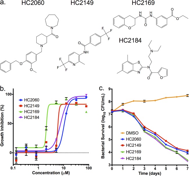
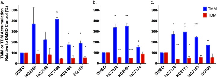
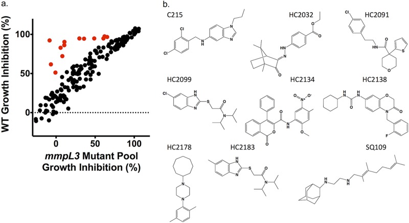
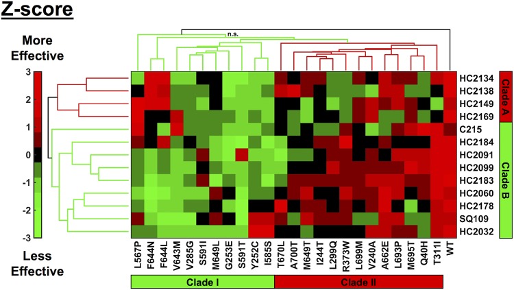
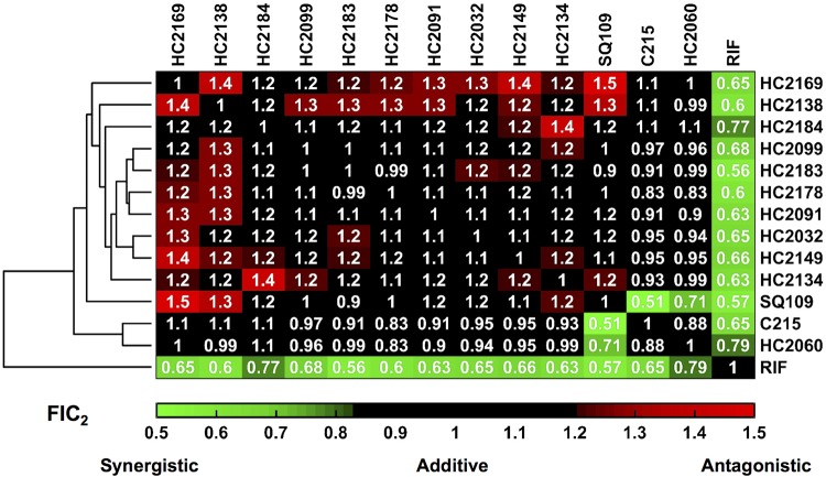
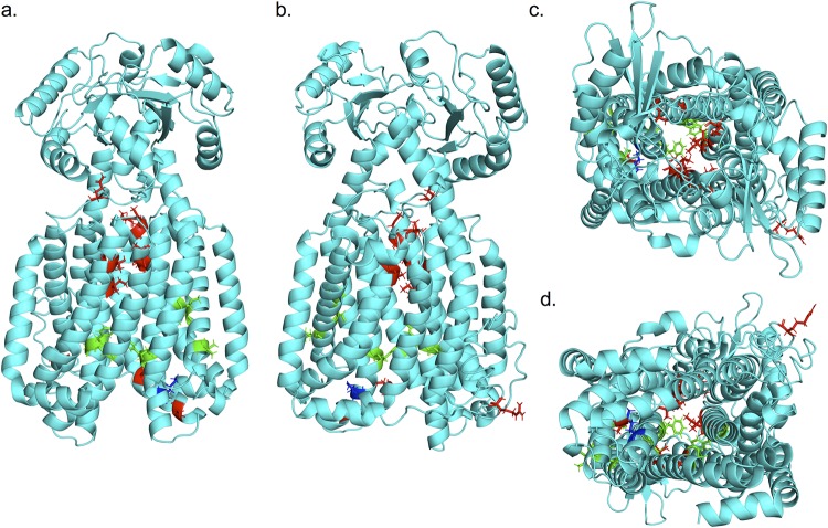
The Mycobacterium tuberculosis mycolate flippase MmpL3 has been the proposed target for multiple inhibitors with diverse chemical scaffolds. This diversity in chemical scaffolds has made it difficult to predict compounds that inhibit MmpL3 without whole-genome sequencing of isolated resistant mutants. Here, we describe the identification of four new inhibitors that select for resistance mutations in mmpL3. Using these resistant mutants, we conducted a targeted whole-cell phenotypic screen of 163 novel M. tuberculosis growth inhibitors for differential growth inhibition of wild-type M. tuberculosis compared to the growth of a pool of 24 unique mmpL3 mutants. The screen successfully identified six additional putative MmpL3 inhibitors. The compounds were bactericidal both in vitro and against intracellular M. tuberculosisM. tuberculosis cells treated with these compounds were shown to accumulate trehalose monomycolates, have reduced levels of trehalose dimycolate, and displace an MmpL3-specific probe, supporting MmpL3 as the target. The inhibitors were mycobacterium specific, with several also showing activity against the nontuberculous mycobacterial species M. abscessus Cluster analysis of cross-resistance profiles generated by dose-response experiments for each combination of 13 MmpL3 inhibitors against each of the 24 mmpL3 mutants defined two clades of inhibitors and two clades of mmpL3 mutants. Pairwise combination studies of the inhibitors revealed interactions that were specific to the clades identified in the cross-resistance profiling. Additionally, modeling of resistance-conferring substitutions to the MmpL3 crystal structure revealed clade-specific localization of the residues to specific domains of MmpL3, with the clades showing differential resistance. Several compounds exhibited high solubility and stability in microsomes and low cytotoxicity in macrophages, supporting their further development. The combined study of multiple mutants and novel compounds provides new insights into structure-function interactions of MmpL3 and small-molecule inhibitors.

Keywords: Mycobacterium tuberculosis; antimicrobials; cell envelope; mechanisms of resistance; phenotypic screening.


Figures

FIG 1
FIG 1
Four compounds inhibit M. tuberculosis growth in a dose- and time-dependent manner. (a) Structures of HC2060, HC2149, HC2169, and HC2184. (b) Inhibition of M. tuberculosis growth in a dose-dependent manner. (c) Killing of M. tuberculosis in a time-dependent manner when treated with the inhibitors at 20 μM. Error bars indicate the standard deviations from the mean values. Experiments were conducted in biological triplicates.
FIG 2
FIG 2
Modulation of TMM and TDM accumulation. (a) Whole-cell 14C-lipids from M. tuberculosis cells treated with 20 μM HC2060, HC2149, HC2169, and HC2184 show increased levels of TMM and decreased levels of TDM. (b and c) Whole-cell 14C-lipids from M. tuberculosis cells treated with a concentration of 20 μM of the six inhibitors identified by the targeted phenotypic screen show increased levels of TMM and decreased levels of TDM. Experiments were conducted in biological duplicates. In both experiments, M. tuberculosis samples were treated with DMSO or 20 μM SQ109 as controls. Error bars indicate the standard deviations. *, P < 0.05; **, <0.005; ***, <0.001; ♦, value that just missed the cut off (P = 0.07 compared to TMM level after HC2134 treatment). The results for HC2060 and HC2149 missed the significance cutoff, but this may be due to the high variability in replicates, as there was a >2-fold difference for HC2060 and HC2149.
FIG 3
FIG 3
A targeted whole-cell phenotypic screen identifies six new MmpL3 inhibitors. (a) Results of a direct head-to-head comparison of percentages of growth inhibition of WT M. tuberculosis and a pooled mmpL3 mutant population treated with 163 compounds at 20 μM. Additional treatments included 0.5 μM BDQ, CLO, INH, PAS, or SQ109 or 0.03% H2O2. Examples of hit compounds with reduced activity in the MmpL3 mutant pool are shown in red. (b) Structures of the confirmed hit compounds from the screen, including six new compounds, HC2032, HC2099, HC2134, HC2138, HC2178, and HC2183. Previously described compounds include C215, HC2091, and SQ109.
FIG 4
FIG 4
Flow cytometry-based competition binding assay using intact M. smegmatis cells expressing M. tuberculosis MmpL3 (MmpL3tb). The assay was performed in an M. smegmatis mmpL3 deletion mutant expressing the wild-type mmpL3tb gene (M. smegmatis MsmgΔmmpL3/pMVGH1-mmpL3tb). Cells were labeled with 4 mM North 114 and subsequently treated with increasing concentrations of the inhibitors. Shown on the y axis are the mean fluorescence intensities (MFI) of the bacilli from each treatment group expressed relative to the MFI of bacilli not treated with any inhibitor (relative fluorescence intensity [RFI] arbitrarily set to 1). MFIs were determined by analyzing 10,000 bacilli under each condition. The data reported are mean values ± standard deviations of technical duplicates. Replicate samples were analyzed by t test. *, P ≤ 0.05; ♦, P ≤ 0.1.
FIG 5
FIG 5
Cross-resistance profiling identifies clustering of compounds and mutations. Cluster analysis of cross-resistance profiling of 24 mmpL3 strains treated with each of the 13 MmpL3 inhibitors normalized by Z-scoring by treatment. Compounds clustered into two clades: clade A and clade B. Mutant strains, denoted by amino acid substitution, clustered into two clades: clade I and clade II. Colors are based on Z-score normalization of treatment; green indicates when treatments were less effective than the average, and red indicates when treatments were more effective than the average. Black (n.s., not significant) indicates a branch where the approximate unbiased (AU) value was <75. All other branches were significant based on bootstrap AU values of >75.
FIG 6
FIG 6
DiaMOND analysis identifies additive, synergistic, and antagonistic inhibitor interactions. Hierarchical cluster analysis of DiaMOND-based pairwise inhibitor interactions of all combinations of MmpL3 inhibitors and RIF identifies additive (FIC2 of 0.82 to 1.18), antagonistic (FIC2 > 1.18), and synergistic (FIC2 < 0.82) interactions.
FIG 7
FIG 7
Mutation substitutions cluster according to cross-resistance clades. (a to d) Front, back, top, and bottom views, respectively, of an I-TASSER-predicted structure of M. tuberculosis MmpL3 based on M. smegmatis MmpL3 structure (PDB code 6AJH). Substitutions conferred by mutations in mmpL3 are indicated. Substitutions are colored based on clade from cross-resistance profiling as follows: green, clade I substitutions; red, clade II substitutions; blue, M649, which fell into both clades depending on the substitution. The model shows a truncated version (732/944 aa) of the MmpL3 protein lacking the C-terminal tail.

References

    1. Grzegorzewicz AE, Pham H, Gundi VA, Scherman MS, North EJ, Hess T, Jones V, Gruppo V, Born SE, Kordulakova J, Chavadi SS, Morisseau C, Lenaerts AJ, Lee RE, McNeil MR, Jackson M. 2012. Inhibition of mycolic acid transport across the Mycobacterium tuberculosis plasma membrane. Nat Chem Biol 8:334–341. doi: 10.1038/nchembio.794. - DOI - PMC - PubMed
    1. La Rosa V, Poce G, Canseco JO, Buroni S, Pasca MR, Biava M, Raju RM, Porretta GC, Alfonso S, Battilocchio C, Javid B, Sorrentino F, Ioerger TR, Sacchettini JC, Manetti F, Botta M, De Logu A, Rubin EJ, De Rossi E. 2012. MmpL3 is the cellular target of the antitubercular pyrrole derivative BM212. Antimicrob Agents Chemother 56:324–331. doi: 10.1128/AAC.05270-11. - DOI - PMC - PubMed
    1. Stanley SA, Grant SS, Kawate T, Iwase N, Shimizu M, Wivagg C, Silvis M, Kazyanskaya E, Aquadro J, Golas A, Fitzgerald M, Dai H, Zhang L, Hung DT. 2012. Identification of novel inhibitors of M. tuberculosis growth using whole cell based high-throughput screening. ACS Chem Biol 7:1377–1384. doi: 10.1021/cb300151m. - DOI - PMC - PubMed
    1. Tahlan K, Wilson R, Kastrinsky DB, Arora K, Nair V, Fischer E, Barnes SW, Walker JR, Alland D, Barry CE III, Boshoff HI. 2012. SQ109 targets MmpL3, a membrane transporter of trehalose monomycolate involved in mycolic acid donation to the cell wall core of Mycobacterium tuberculosis. Antimicrob Agents Chemother 56:1797–1809. doi: 10.1128/AAC.05708-11. - DOI - PMC - PubMed
    1. Lun S, Guo H, Onajole OK, Pieroni M, Gunosewoyo H, Chen G, Tipparaju SK, Ammerman NC, Kozikowski AP, Bishai WR. 2013. Indoleamides are active against drug-resistant Mycobacterium tuberculosis. Nat Commun 4:2907. doi: 10.1038/ncomms3907. - DOI - PMC - PubMed

Publication types

MeSH terms

LinkOut - more resources