. 2019 Aug 12;9(1):11601.
doi: 10.1038/s41598-019-48014-6.

Autosomal Dominant Tubulointerstitial Kidney Disease with Adult Onset due to a Novel Renin Mutation Mapping in the Mature Protein

Affiliations

Autosomal Dominant Tubulointerstitial Kidney Disease with Adult Onset due to a Novel Renin Mutation Mapping in the Mature Protein

Céline Schaeffer et al. Sci Rep. .

Abstract

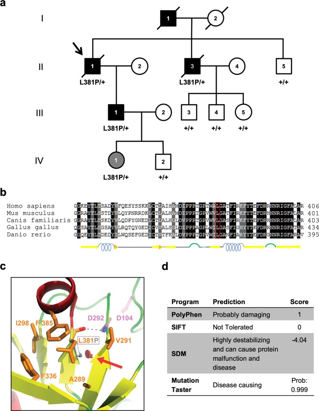
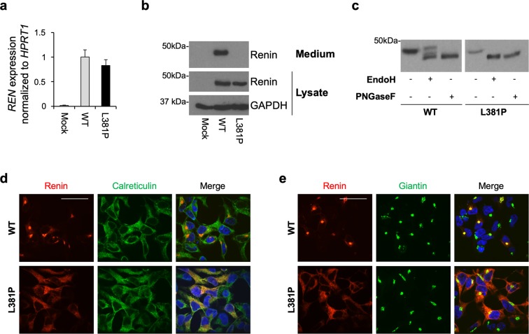
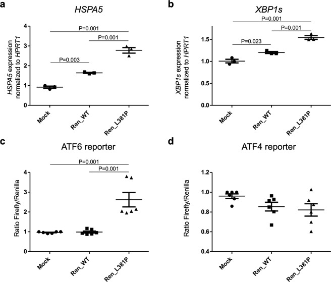
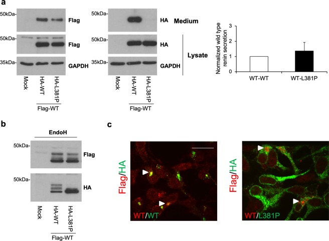
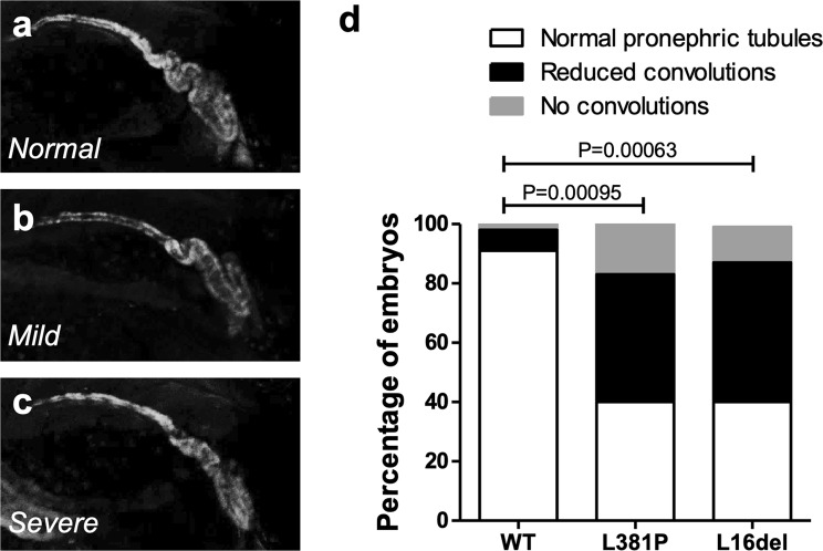
Autosomal dominant tubulointerstitial kidney disease (ADTKD) is a genetically heterogeneous renal disorder leading to progressive loss of renal function. ADTKD-REN is due to rare mutations in renin, all localized in the protein leader peptide and affecting its co-translational insertion in the endoplasmic reticulum (ER). Through exome sequencing in an adult-onset ADTKD family we identified a new renin variant, p.L381P, mapping in the mature protein. To assess its pathogenicity, we combined genetic data, computational and predictive analysis and functional studies. The L381P substitution affects an evolutionary conserved residue, co-segregates with renal disease, is not found in population databases and is predicted to be deleterious by in silico tools and by structural modelling. Expression of the L381P variant leads to its ER retention and induction of the Unfolded Protein Response in cell models and to defective pronephros development in zebrafish. Our work shows that REN mutations outside of renin leader peptide can cause ADTKD and delineates an adult form of ADTKD-REN, a condition which has usually its onset in childhood. This has implications for the molecular diagnosis and the estimated prevalence of the disease and points at ER homeostasis as a common pathway affected in ADTKD-REN, and possibly more generally in ADTKD.


Conflict of interest statement

The authors declare no competing interests.

Figures

Figure 1
Figure 1
Identification of L381P renin variant in an ADTKD family. (a) Pedigree chart of the ADTKD family segregating the renin p.L381P variant. Index case (arrow): II-1. Black symbols denote clinically affected individuals, open symbols denote clinically unaffected individuals. Corresponding REN genotype is provided for each tested individual. (b) Alignment of renin sequence from human, mouse, dog, chicken and zebrafish performed with the Sequence Manipulation Suite. Identical or similar residues are shown with a black and grey background respectively. Predicted secondary structure is shown below. (c) Detail of the human renin structure, shown in cartoon representation and colored by secondary structure (red: α-helices; yellow: β-strands; green: loops). L381 and nearby conserved hydrophobic residues (orange), as well as the two catalytic aspartates (magenta), are depicted as sticks; a red arrow indicates the steric clash (red discs) that would be introduced upon mutation of L381 to proline (grey stick). The main-chain hydrogen bond between L381 and D292 is represented by a dashed black line. The figure was made with PyMOL (Schrödinger LLC). (d) Predicted effect of the renin L381P variant, as assessed by using PolyPhen-2, Sorting Intolerant From Tolerant (SIFT), Site Directed Mutator (SDM) and Mutation Taster.
Figure 2
Figure 2
The L381P renin isoform is retained in the ER. (a) Renin expression in stably transfected HEK cells assessed by real-time RT-qPCR. Expression is normalized to HPRT1. Data are expressed as mean ± s.d. (n = 3 independent experiments). (b) Western blot analysis showing renin expression in cell lysate and conditioned medium. GAPDH is shown as a loading control. No secreted renin can be detected for the L381P isoform. (c) Western blot analysis showing renin in HEK lysates after treatment with EndoH or PNGaseF. Full-length blots (b,c) are presented in Supplementary Fig. S1. (d) Immunofluorescence analysis showing renin (red) and calreticulin (ER marker, green) and merged pictures with nuclei (dapi, blue). Bar = 40 μm. The variant L381P shows co-localization with calreticulin demonstrating its ER retention. (e) Immunofluorescence analysis showing renin (red) and giantin (Golgi marker, green) and merged pictures with nuclei (dapi, blue). Bar = 40 μm. Co-localization with giantin is seen for the wild type isoform only, demonstrating its trafficking along the secretory pathway.
Figure 3
Figure 3
Expression of L381P renin isoform induces the UPR. (a) HSPA5 and (b) XBP1s expression analysis by real-time RT-qPCR. Gene expression is normalized to HPRT1. Expression of L381P isoform in HEK cells induces expression of HSPA5 (BiP) and spliced XBP1, markers of ER stress and UPR respectively. Data are expressed as mean ± s.d. (n = 3 independent experiments) (one way ANOVA, P = 1.78 ×    10−5 and P = 1 × 10−4 for HSPA5 and spliced XBP1 respectively, followed by Tukey Honest Significant Difference (HSD) post hoc test). (c) ATF6 activation assessed through the use of a luciferase-based, ATF6 reporter construct. ATF6 activation is observed upon expression of the L381P renin isoform. Data are expressed as mean ± s.d. (n = 6 independent experiments) (one way ANOVA, P = 5.78 × 10−5, followed by Tukey HSD post hoc test). (d) ATF4 activity, assessed through the use of a luciferase-based reporter construct. No increase of luciferase activity is observed in cells expressing the L381P renin isoform compared to mock or cells expressing wild type renin. Data are expressed as mean ± s.d. (n = 6 independent experiments) (one way ANOVA, P = 0.1175).
Figure 4
Figure 4
The L381P renin isoform does not exert a dominant negative effect on trafficking of wild type renin. (a) Western blot analysis showing Flag- and HA-tagged renin isoforms in lysate and conditioned medium of stably transfected HEK cells. GAPDH is shown as a loading control. Quantification of secreted Flag-tagged wild type isoform normalized to Flag-tagged wild type protein present in cell lysate is shown. Data are expressed as mean ± s.d. (n = 3 independent experiments). (b) Western blot analysis showing Flag- and HA-tagged renin in HEK lysates after treatment with EndoH. Full-length blots (a,b) are presented in Supplementary Fig. S3. (c) Immunofluorescence analysis showing merged picture of Flag- and HA-tagged renin in red and green respectively. Bar = 40 μm. Cellular localization of wild type renin is not affected by co-expression with ER-retained L381P isoform.
Figure 5
Figure 5
Expression of L381P renin variant induces alteration in the pronephros of zebrafish embryos. The pronephros of 5 dpf zebrafish embryos is labelled by reporter transgene EGFP (line ia20Tg). (a–c) Embryos injected with wild type (WT), L381P and L16del renin mRNAs show three different phenotypes: normal convoluted pronephric tubules; tubules with reduced convolutions (mild phenotype); absence of tubular convolution (severe phenotype). (d) Histograms showing the percentage of embryos with normal pronephros, or mild or severe pronephric alterations. Samples injected with renin mutants L381P (n = 112) and L16del (n = 122) show significantly increased frequency of altered pronephric tubules relative to wild type (n = 183) (chi-square analysis).

References

    1. Bleyer AJ, Kidd K, Zivna M, Kmoch S. Autosomal Dominant Tubulointerstitial Kidney Disease. Adv Chronic Kidney Dis. 2017;24:86–93. doi: 10.1053/j.ackd.2016.11.012. - DOI - PMC - PubMed
    1. Gast C, et al. Autosomal dominant tubulointerstitial kidney disease-UMOD is the most frequent non polycystic genetic kidney disease. BMC Nephrol. 2018;19:301. doi: 10.1186/s12882-018-1107-y. - DOI - PMC - PubMed
    1. Knaup KX, et al. Biallelic Expression of Mucin-1 in Autosomal Dominant Tubulointerstitial Kidney Disease: Implications for Nongenetic Disease Recognition. J Am Soc Nephrol. 2018;29:2298–2309. doi: 10.1681/ASN.2018030245. - DOI - PMC - PubMed
    1. Zivna M, et al. Dominant renin gene mutations associated with early-onset hyperuricemia, anemia, and chronic kidney failure. Am J Hum Genet. 2009;85:204–213. doi: 10.1016/j.ajhg.2009.07.010. - DOI - PMC - PubMed
    1. Bleyer AJ, et al. Clinical and molecular characterization of a family with a dominant renin gene mutation and response to treatment with fludrocortisone. Clin Nephrol. 2010;74:411–422. doi: 10.5414/CNP74411. - DOI - PMC - PubMed

Publication types

LinkOut - more resources