. 2019 Aug 12;9(1):11679.
doi: 10.1038/s41598-019-48212-2.

HER2, chromosome 17 polysomy and DNA ploidy status in breast cancer; a translational study

Affiliations

HER2, chromosome 17 polysomy and DNA ploidy status in breast cancer; a translational study

Altuna Halilovic et al. Sci Rep. .

Abstract

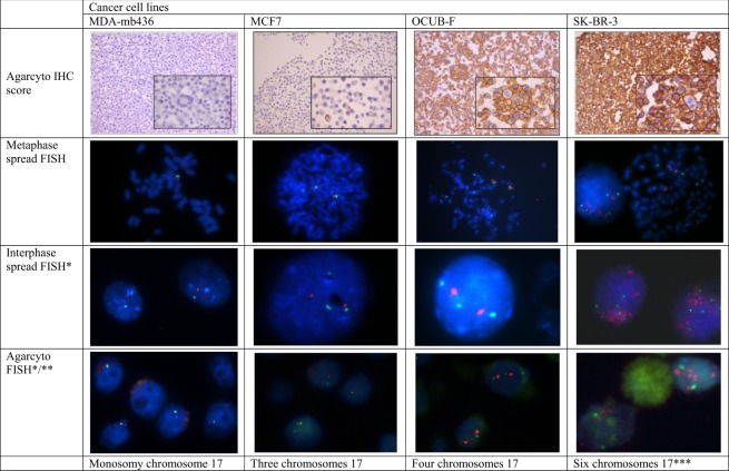
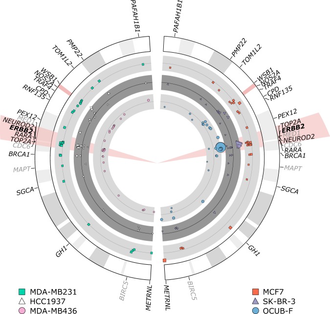
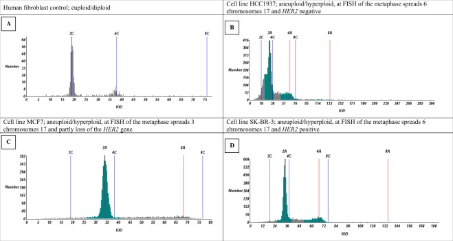
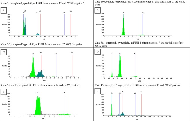
Breast cancer treatment depends on human epidermal growth factor receptor-2 (HER2) status, which is often determined using dual probe fluorescence in situ hybridisation (FISH). Hereby, also loss and gain of the centromere of chromosome 17 (CEP17) can be observed (HER2 is located on chromosome 17). CEP17 gain can lead to difficulty in interpretation of HER2 status, since this might represent true polysomy. With this study we investigated whether isolated polysomy is present and how this effects HER2 status in six breast cancer cell lines and 97 breast cancer cases, using HER2 FISH and immunohistochemistry, DNA ploidy assessment and multiplex ligation dependent probe amplification. We observed no isolated polysomy of chromosome 17 in any cell line. However, FISH analysis did show CEP17 gain in five of six cell lines, which reflected gains of the whole chromosome in metaphase spreads and aneuploidy with gain of multiple chromosomes in all these cases. In patients' samples, gain of CEP17 indeed correlated with aneuploidy of the tumour (91.1%; p < 0.001). Our results indicate that CEP17 gain is not due to isolated polysomy, but rather due to widespread aneuploidy with gain of multiple chromosomes. As aneuploidy is associated with poor clinical outcome, irrespective of tumour grade, this could improve future therapeutic decision making.


Conflict of interest statement

The authors declare no competing interests.

Figures

Figure 1
Figure 1
Examples of HER2 IHC of agarcyto slides and HER2/CEP17 FISH results on metaphase spreads, interphase nuclei, and agarcyto slides. Due to focusing (*) or cutting artefacts (**), not all of the spots of CEP17 (green) or the HER2 gene (red) may be visible. ***HER2 gene amplified.
Figure 2
Figure 2
MLPA results of chromosome 17 of all cell lines used. Cell lines MDA-MB231, HCC1937, MCF7, SK-BR-3, OCUB-F and MDA-MB436 are shown. Probemix P004-B1 and P078-B1 (greytone) were used on all cell lines. The HER2 gene is highlighted in red. The size of the used pictograms corresponds to the reach of the MLPA results (bigger size correlates to higher reach). Results between the probemixes were concordant. The P078-B1 breast tumour probemix also contained probes for genes located on other chromosomes relevant for breast carcinoma and reference genes. In these genes no polysomy or loss or amplification could be detected (not shown in the figure). Negative controls used, were nicely oriented around 1,00 (not shown). The MLPA results with probemix P004-B1 and P078-B1 (greytone) of cell lines MDA-MB231 and HCC1937 show no polysomy or loss or amplification of HER2. MCF7 shows partial loss of the HER2 gene, corresponding to FISH results (Table 1). SK-BR-3 shows amplification of the HER2 gene corresponding to IHC results and FISH results (Fig. 1). OCUB-F shows amplification of the HER2 gene corresponding to the IHC and FISH results (Table 1). MDA-MB436 shows partial loss of all tested genes on chromosome 17, concordant with monosomy, as was seen with FISH (Fig. 1).
Figure 3
Figure 3
Cell line examples of DNA histograms of agarcyto material. IOD: integrated optical density.
Figure 4
Figure 4
Breast cancer examples of DNA histograms. IOD: integrated optical density. *Probably hypoploidy although morphologically it was not possible to differentiate tumour nuclei from normal nuclei.

References

    1. Piccart-Gebhart MJ, et al. Trastuzumab after adjuvant chemotherapy in HER2-positive breast cancer. N Engl J Med. 2005;353:1659–1672. doi: 10.1056/NEJMoa052306. - DOI - PubMed
    1. Romond EH, et al. Trastuzumab plus adjuvant chemotherapy for operable HER2-positive breast cancer. N Engl J Med. 2005;353:1673–1684. doi: 10.1056/NEJMoa052122. - DOI - PubMed
    1. Smith I, et al. 2-year follow-up of trastuzumab after adjuvant chemotherapy in HER2-positive breast cancer: a randomised controlled trial. Lancet. 2007;369:29–36. doi: 10.1016/S0140-6736(07)60028-2. - DOI - PubMed
    1. Joensuu H, et al. Fluorouracil, epirubicin, and cyclophosphamide with either docetaxel or vinorelbine, with or without trastuzumab, as adjuvant treatments of breast cancer: final results of the FinHer Trial. J Clin Oncol. 2009;27:5685–5692. doi: 10.1200/JCO.2008.21.4577. - DOI - PubMed
    1. Chan A. A review of the use of trastuzumab (Herceptin) plus vinorelbine in metastatic breast cancer. Ann Oncol. 2007;18:1152–1158. doi: 10.1093/annonc/mdl476. - DOI - PubMed

MeSH terms