

doi: 10.1038/s41598-019-47660-0.
Role of Hydrogen Bonding in Green Fluorescent Protein-like Chromophore Emission
Affiliations
- PMID: 31406231
- PMCID: PMC6690883
- DOI: 10.1038/s41598-019-47660-0
Item in Clipboard
Role of Hydrogen Bonding in Green Fluorescent Protein-like Chromophore Emission
Sci Rep.
.
Abstract
The fluorescence emission from green fluorescent protein (GFP) is known to be heavily influenced by hydrogen bonding between the core fluorophore and the surrounding side chains or water molecules. Yet how to utilize this feature for modulating the fluorescence of GFP chromophore or GFP-like fluorophore still remains elusive. Here we present theoretical calculations to predict how hydrogen bonding could influence the excited states of the GFP-like fluorophores. These studies provide both a new perspective for understanding the photophysical properties of GFP as well as a solid basis for the rational design of GFP-based fluorophores.
Conflict of interest statement
The authors declare no competing interests.
Figures

Photo absorption and emission process of the modified GFP chromophore. (a) Chemical structures of the GFP chromophore and (b) the C=C double bond-modified GFP chromophore. (c) The computed absorption spectrum together with the lowest photo-emission peak (d) of the modified chromophore. The calculated transitions between frontier orbitals for the photo absorption (e) and emission (f) of the modified chromophore in aqueous solution. The blue, gray, white and red balls represent N, C, H and O atoms, respectively. f stands for oscillator strength.
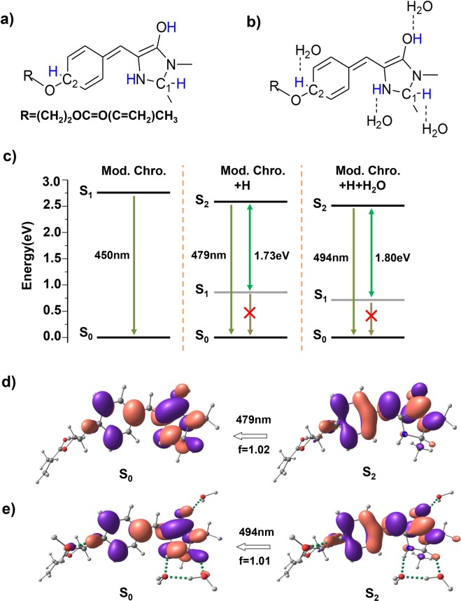
Fluorescent redshift of the modified chromophore with hydrogen bonding network consisting of hydrogen and water. Molecular configurations of the modified chromophore with hydrogen in the (a) absence and (b) presence of water molecules, added hydrogen atom are highlighted in blue. (c) The computed electronic energy levels and photo-emission transitions of the two excited-state structures and the original modified chromophore. The orbital transitions for the calculated photo-emission peak of the chromophore with hydrogen in the (d) absence and (e) presence of water molecules in aqueous solution. The green dotted bonds denote hydrogen bonding.
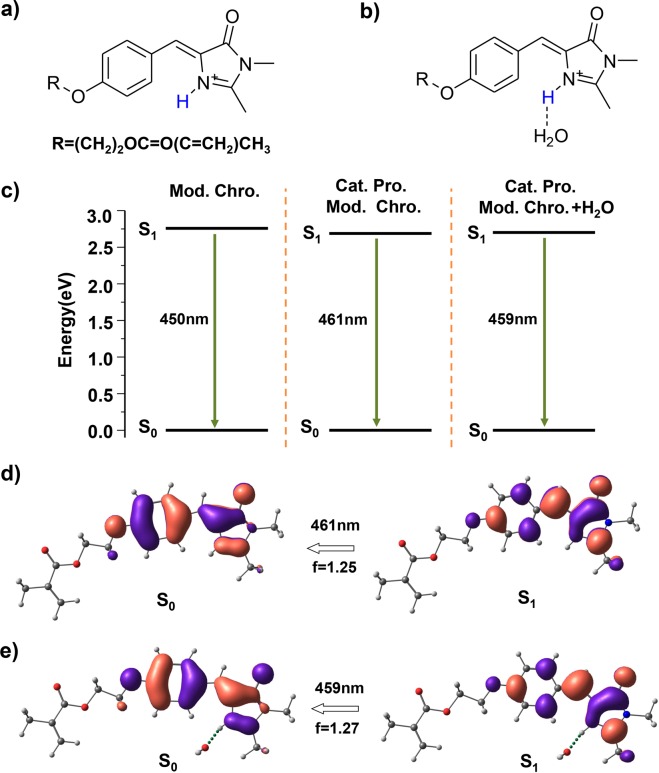
Optical properties of the cationic protonated modified chromophore and that with water molecule. Molecular configurations of the monocationic protonated modified structure in the (a) absence and (b) presence of water molecule, added protons are highlighted in blue. (c) The computed electronic energy levels and photo-emission transitions of these two excited-state structures and the original modified chromophore. The calculated transitions between frontier orbitals for the photo-emission peak of the protonated cation in the (d) absence and (e) presence of a water molecule.
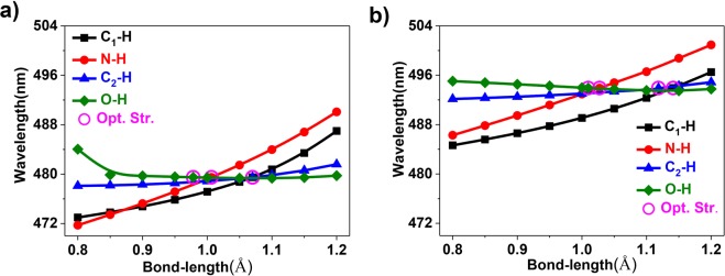
Relationship between the emission wavelengths and relevant bond lengths. The calculated emission maximum as a function of selected bond lengths for the modified chromophore with hydrogen in the (a) absence and (b) presence of water molecules in aqueous solution.
References
Publication types
MeSH terms
Substances
LinkOut - more resources
Full Text Sources
Molecular Biology Databases
