. 2019 Aug 12;9(1):11717.
doi: 10.1038/s41598-019-48282-2.

Atorvastatin increases the production of proinflammatory cytokines and decreases the survival of Escherichia coli-infected mice

Affiliations

Atorvastatin increases the production of proinflammatory cytokines and decreases the survival of Escherichia coli-infected mice

Hadi M Hussein et al. Sci Rep. .

Abstract

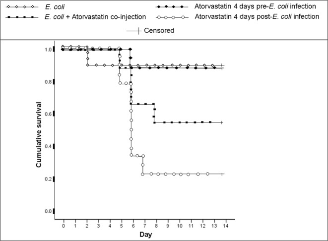
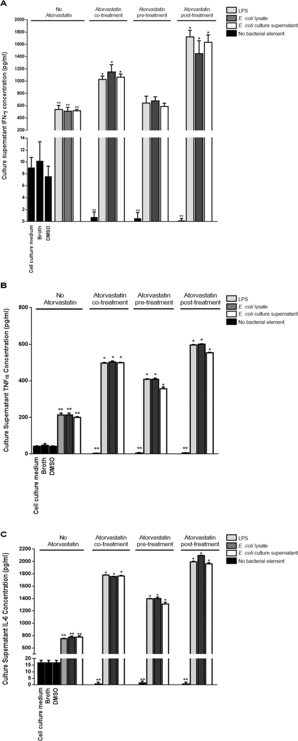
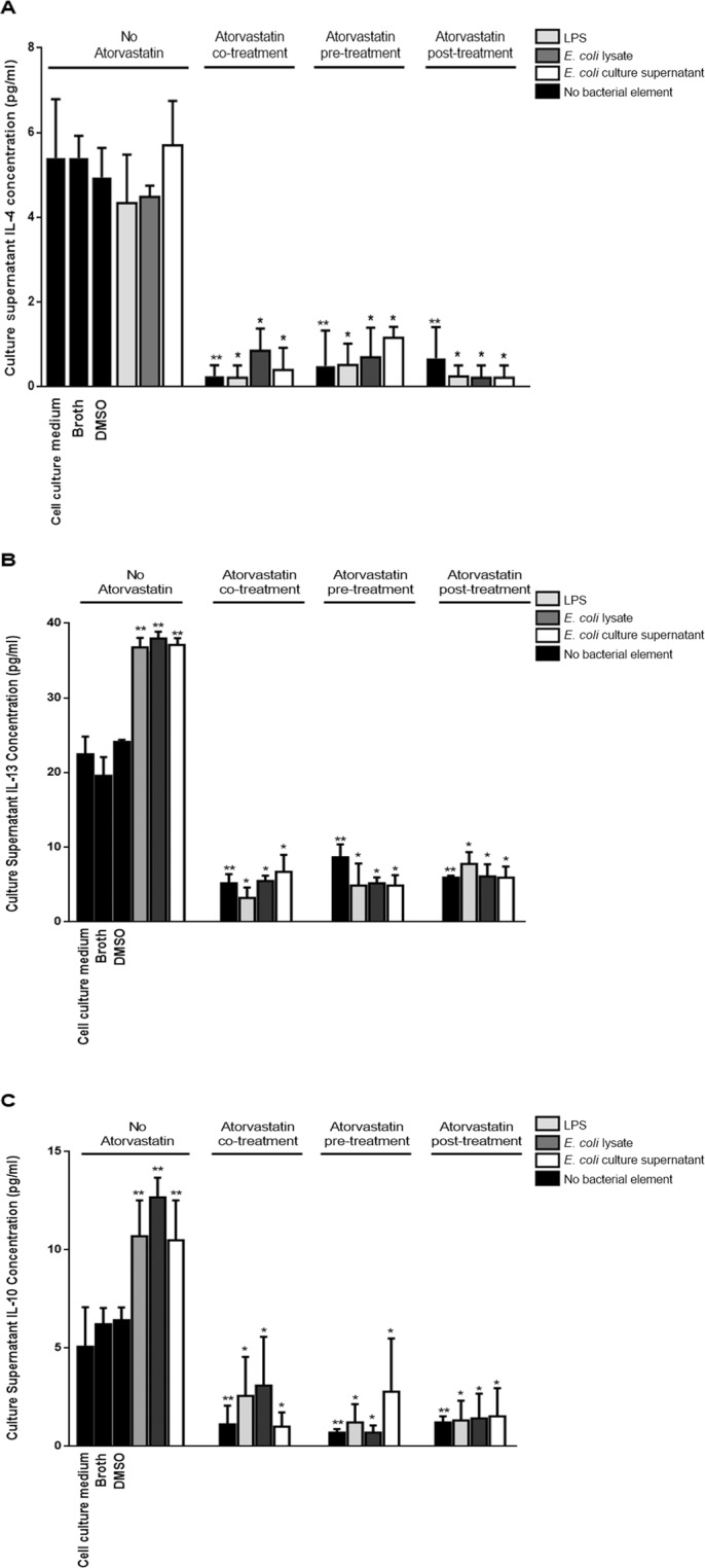
To assess whether the immunosuppressive effects of atorvastatin outweigh its antibacterial ones in an infection, mice were infected with Escherichia coli and administered atorvastatin; survival rates were then monitored. Mice treated with atorvastatin post-infection showed a remarkable decrease in their survival rate. On the other hand, the higher the level of serum IFN-γ in the infected mice treated with atorvastatin, the lower was the survival rate. Levels of IL-4 were markedly depressed in all groups infected with E. coli and treated with atorvastatin. Since atorvastatin inhibits IFN-γ expression in the absence of bacterial infection, we examined whether bacterial lipopolysaccharide (LPS) was the element capable of overriding this inhibition. Mouse peripheral blood mononuclear cells were treated with atorvastatin and lipopolysaccharide ex vivo then proinflammatory (IFN-γ, TNFα, IL-6) and prohumoral/regulatory (IL-4, IL-13, IL-10) cytokine levels were analyzed in culture supernatants. While proinflammatory cytokine levels were decreased upon treatment with atorvastatin alone, their levels were markedly elevated by treatment with LPS, bacterial lysate or bacterial culture supernatant. On the other hand, atorvastatin exerted an inhibitory effect on production of the prohumoral/regulatory cytokines. Our data indicates that any consideration for statins as antimicrobial treatment should assess the possible adverse outcomes.


Conflict of interest statement

The authors declare no competing interests.

Figures

Figure 1
Figure 1
Effect of atorvastatin treatment on the survival of E. coli-infected mice. BALB/c mice (9 per group) were intraperitoneally injected with E. coli and treated either with a single co-injection of atorvastatin, a 4-day pre-infection or a 4-day post-infection atorvastatin regimen. A group infected with E. coli but not treated with atorvastatin was also included. Day 0 indicates the day of infection. Mice were monitored for 14 days after infection. The remaining number of mice at the end of the monitoring period was 2 in the group treated with atorvastatin for 4 days post-infection, 4 in the group administered a single dose of atorvastatin upon infection, and 8 in both the group treated with atorvastatin for 4 days pre-infection or infected but not treated with atorvastatin.
Figure 2
Figure 2
Serum IFN-γ levels in mice infected with E. coli and treated with atorvastatin. BALB/c mice (9 per group) received an intraperitoneal injection E. coli and were treated either with a single co-injection of atorvastatin, a 4-day pre-infection or a 4-day post-infection atorvastatin regimen. A group that was infected with E. coli but not treated with atorvastatin was included as well. Mouse sera were analyzed on day 4 after infection for IFN-γ levels by ELISA. *indicates p < 0.05 compared to the E. coli-infected group.
Figure 3
Figure 3
Serum IL-4 levels in mice infected with E. coli and treated with atorvastatin. BALB/c mice (9 per group) received an intraperitoneal injection E. coli and were treated either with a single co-injection of atorvastatin, a 4-day pre-infection or a 4-day post-infection atorvastatin regimen. A group that was infected with E. coli but not treated with atorvastatin was included as well. Mouse sera were analyzed on day 4 after infection for IL-4 levels by ELISA. *indicates p < 0.05 compared to the E. coli-infected group.
Figure 4
Figure 4
Proinflammatory cytokine levels in culture supernatants from mouse PBMCs incubated with bacterial components and treated with atorvastatin. Mouse PBMCs were cultured for 12 hrs at 37 °C prior to introduction of a bacterial treatment element [Lipopolysaccharide (LPS)/E. coli lysate/E. coli culture supernatant]. Cells were incubated with each bacterial element for another 12 hrs prior to culture supernatant collection for cytokine level analysis by ELISA. Atorvastatin was introduced 4 hrs before, along with or 4 hrs after introduction of the bacterial treatment element. Control treatments consisted of culture medium alone (LPS solvent), DMSO (atorvastatin solvent) or sterile Brain Heart Infusion (BHI) broth; these were added to cells that were cultured for 12 hrs at 37 °C. Atorvastatin control treatments were also included with the statin being added in a timeline similar to that used for the bacterial element-treated cells but in the absence of this latter type of treatment. (A) IFN-γ levels. (B) TNFα levels. (C) IL-6 levels. *indicates p < 0.05 compared to the respective E. coli treatment element in absence of atorvastatin; **indicates p < 0.05 compared to cells mock treated with vehicle (Culture medium for LPS, BHI broth for E.coli lysates or culture supernatant and DMSO for atorvastatin).
Figure 5
Figure 5
Prohumoral/regulatory cytokine levels in culture supernatants from mouse PBMCs incubated with bacterial components and treated with atorvastatin. Mouse PBMCs were cultured for 12 hrs at 37 °C prior to introduction of a bacterial treatment element [Lipopolysaccharide (LPS)/E. coli lysate/E. coli culture supernatant]. Cells were incubated with each bacterial element for another 12 hrs prior to culture supernatant collection for cytokine level analysis by ELISA. Atorvastatin was introduced 4 hrs before, along with or 4 hrs after introduction of the bacterial treatment element. Control treatments consisted of culture medium alone (LPS solvent), DMSO (atorvastatin solvent) or sterile Brain Heart Infusion broth; these were added to cells that were already cultured for 12 hrs at 37 °C. Atorvastatin control treatments were also included with the statin being added in a timeline similar to that used for the bacterial element-treated cells but in the absence of this latter type of treatment. (A) IL-4 levels. (B) IL-13 levels. (C) IL-10 levels. *indicates p < 0.05 compared to the respective E. coli treatment element in absence of atorvastatin; **indicates p < 0.05 compared to cells mock treated with DMSO.

References

    1. Cordle A, Koenigsknecht-Talboo J, Wilkinson B, Limpert A, Landreth G. Mechanisms of statin-mediated inhibition of small G-protein function. J Biol Chem. 2005;280:34202–34209. doi: 10.1074/jbc.M505268200. - DOI - PubMed
    1. Khan MA, et al. Statins impair CD1d-mediated antigen presentation through the inhibition of prenylation. J Immunol. 2009;182:4744–4750. doi: 10.4049/jimmunol.0804311. - DOI - PMC - PubMed
    1. Van Aelst L, D’Souza-Schorey C. Rho GTPases and signaling networks. Genes Dev. 1997;11:2295–2322. doi: 10.1101/gad.11.18.2295. - DOI - PubMed
    1. Pick E. Role of the Rho GTPase Rac in the activation of the phagocyte NADPH oxidase: outsourcing a key task. Small GTPases. 2014;5:e27952. doi: 10.4161/sgtp.27952. - DOI - PMC - PubMed
    1. Hechinger AK, et al. Inhibition of protein geranylgeranylation and farnesylation protects against graft-versus-host disease via effects on CD4 effector T cells. Haematologica. 2013;98:31–40. doi: 10.3324/haematol.2012.065789. - DOI - PMC - PubMed

Publication types

MeSH terms