. 2019 Dec;4(12):2204-2215.
doi: 10.1038/s41564-019-0520-8. Epub 2019 Aug 12.

DABs are inorganic carbon pumps found throughout prokaryotic phyla

Affiliations

DABs are inorganic carbon pumps found throughout prokaryotic phyla

John J Desmarais et al. Nat Microbiol. 2019 Dec.

Abstract

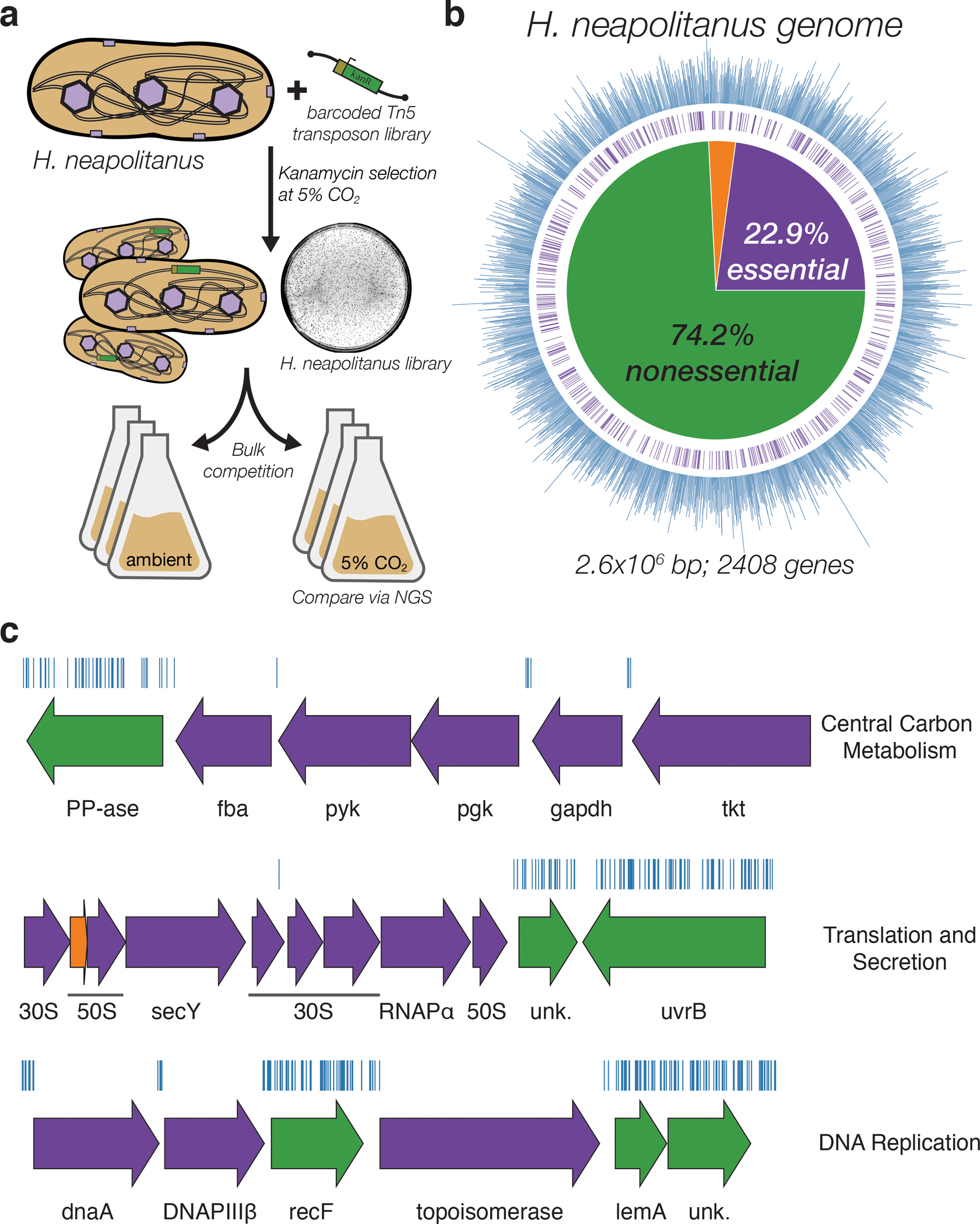
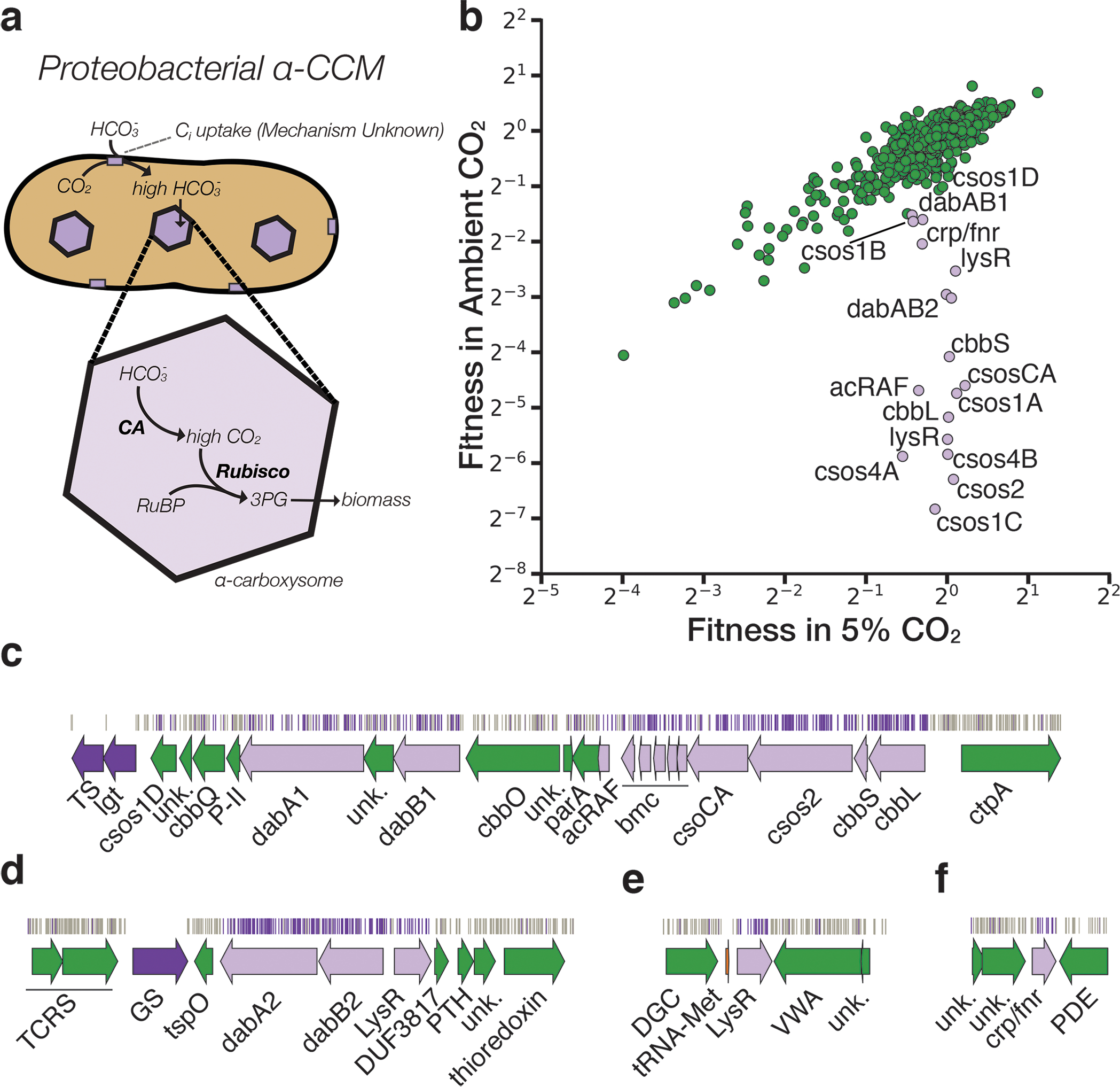
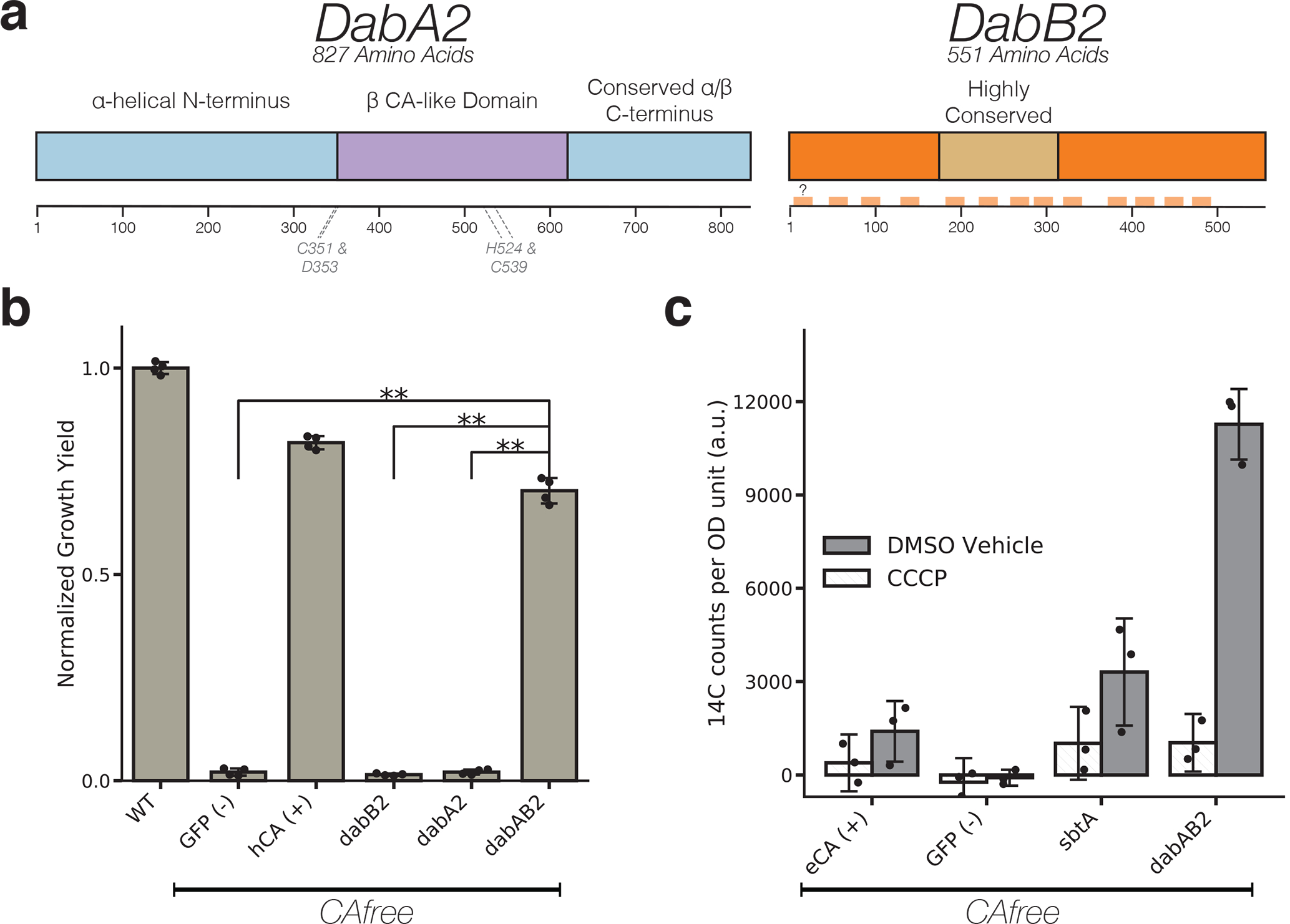
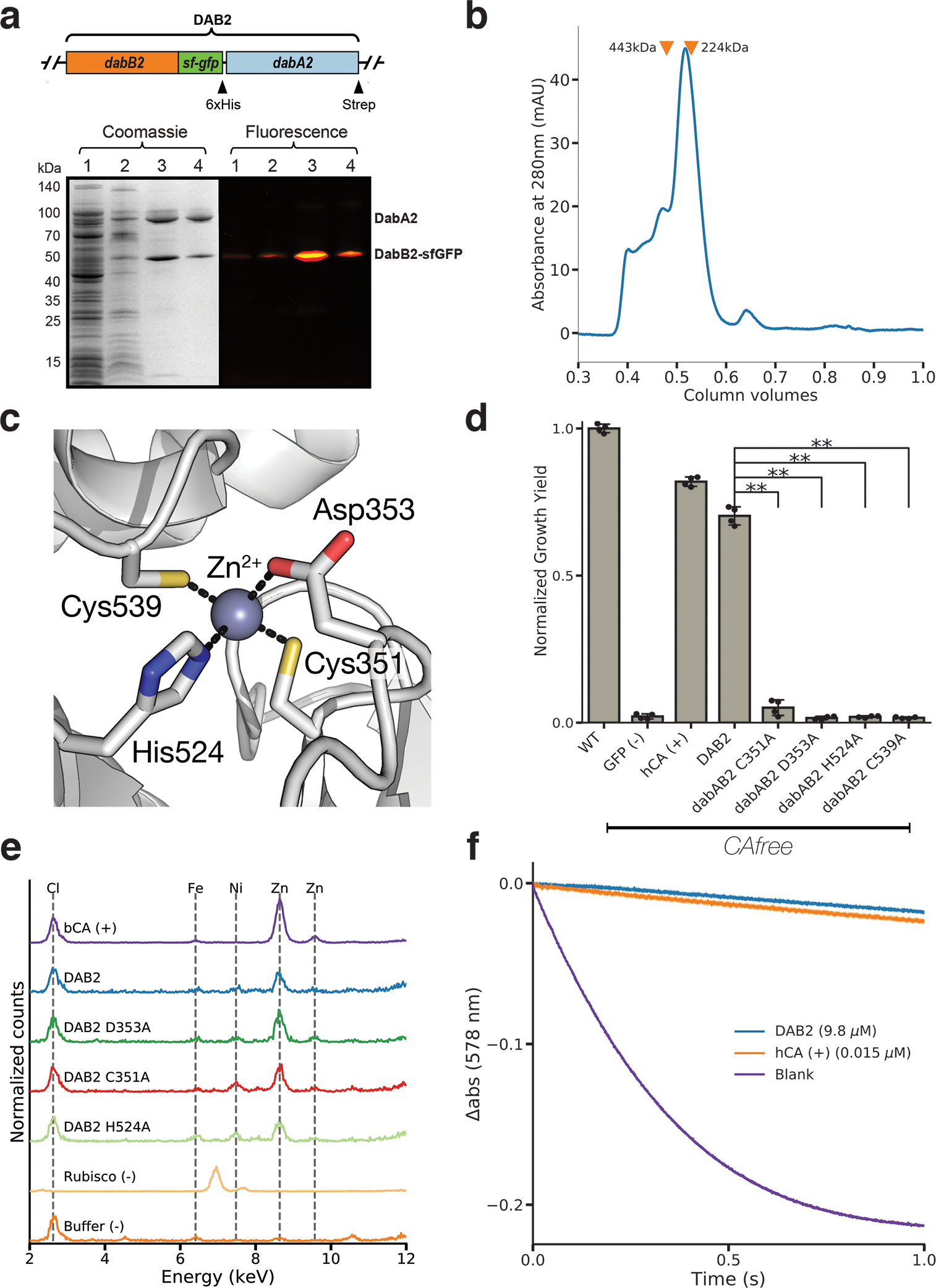
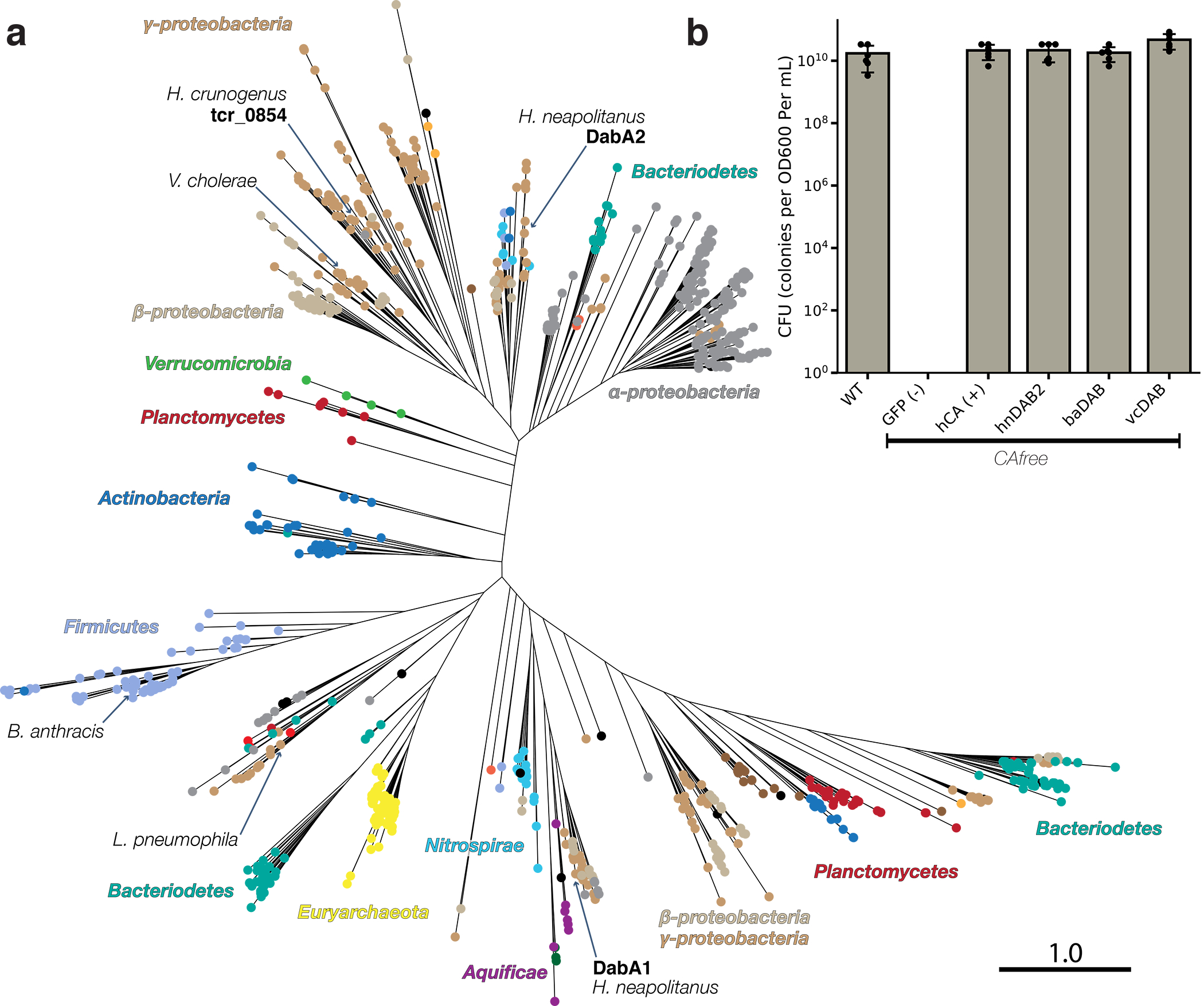
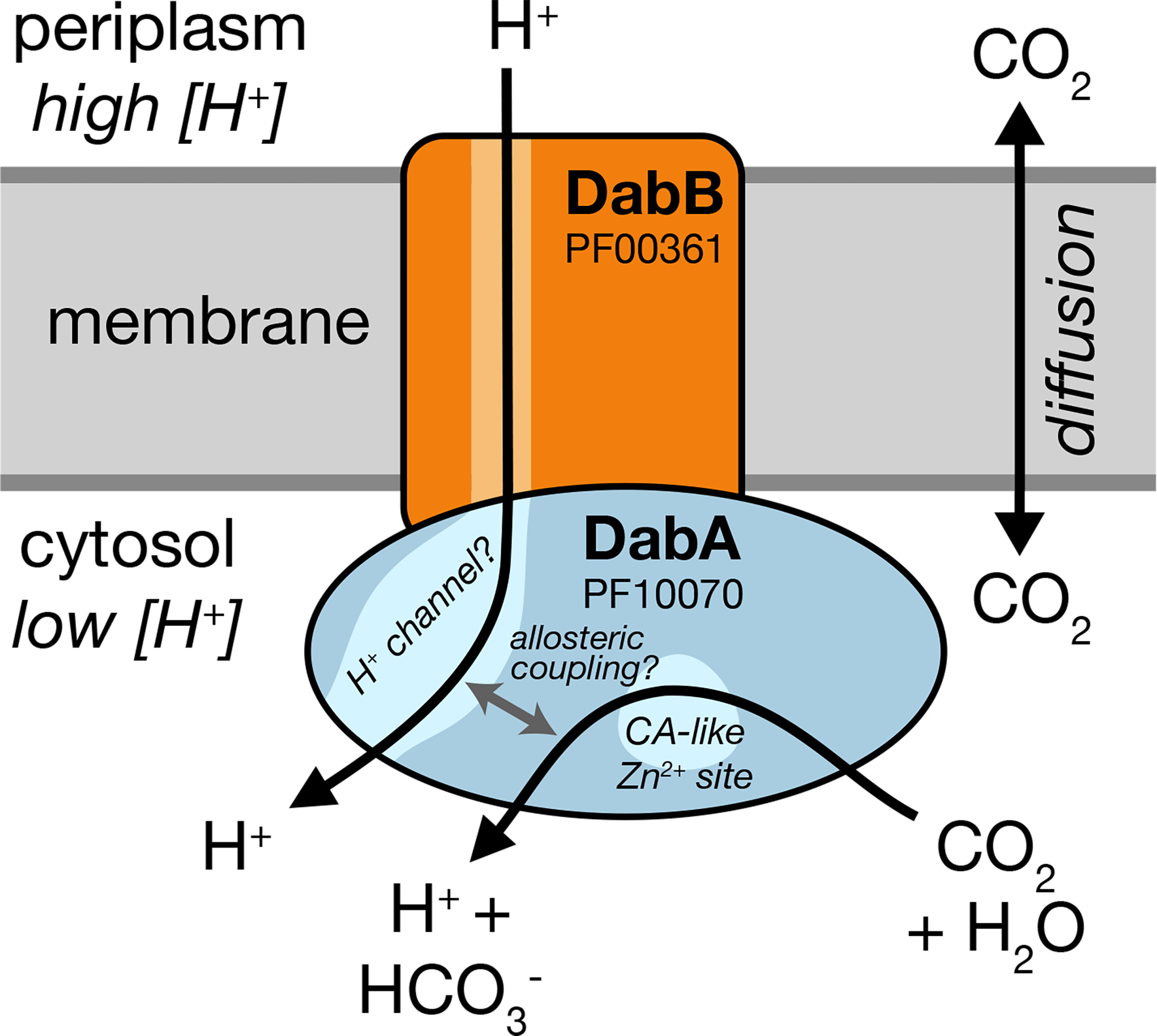
Bacterial autotrophs often rely on CO2 concentrating mechanisms (CCMs) to assimilate carbon. Although many CCM proteins have been identified, a systematic screen of the components of CCMs is lacking. Here, we performed a genome-wide barcoded transposon screen to identify essential and CCM-related genes in the γ-proteobacterium Halothiobacillus neapolitanus. Screening revealed that the CCM comprises at least 17 and probably no more than 25 genes, most of which are encoded in 3 operons. Two of these operons (DAB1 and DAB2) contain a two-gene locus that encodes a domain of unknown function (Pfam: PF10070) and a putative cation transporter (Pfam: PF00361). Physiological and biochemical assays demonstrated that these proteins-which we name DabA and DabB, for DABs accumulate bicarbonate-assemble into a heterodimeric complex, which contains a putative β-carbonic anhydrase-like active site and functions as an energy-coupled inorganic carbon (Ci) pump. Interestingly, DAB operons are found in a diverse range of bacteria and archaea. We demonstrate that functional DABs are present in the human pathogens Bacillus anthracis and Vibrio cholerae. On the basis of these results, we propose that DABs constitute a class of energized Ci pumps and play a critical role in the metabolism of Ci throughout prokaryotic phyla.


Conflict of interest statement

Competing Interests

UC Regents have filed a patent related to this work on which J.J.D., A.F., and D.F.S. are inventors. D.F.S. is a co-founder of Scribe Therapeutics and a scientific advisory board member of Scribe Therapeutics and Mammoth Biosciences. All other authors declare no competing interests.

Figures

Figure 1.
Figure 1.. Transposon mutagenesis reveals the essential gene set of a chemolithoautotrophic organism.
a. Schematic depicting the generation and screening of the RB-TnSeq library. Transposons were inserted into the Hnea genome by conjugation with an E. coli donor strain. The transposon contains a random 20 base pair barcode (yellow) and a kanamycin selection marker (green). Selection for colonies containing insertions was performed in the presence of kanamycin at 5% CO2 and insertions were mapped by sequencing as described in the Methods. Subsequent screens were carried out as bulk competition assays and quantified by BarSeq. b. Insertions and essential genes are well-distributed throughout the Hnea genome. The outer track (blue) is a histogram of the number of barcodes that were mapped to a 1 kb window. The inner track annotates essential genes in purple. The pie chart shows the percentages of the genome called essential (purple), ambiguous (orange), and nonessential (green). c. Representative essential genes and nonessential genes in the Hnea genome. The blue track indicates the presence of an insertion. Genes in purple were called essential and genes in green are nonessential. Genes labeled “unk.” are hypothetical proteins. The first genomic locus contains 5 essential genes involved in glycolysis or the CBB cycle including pyruvate kinase (pyk) and transketolase (tkt). The 8 essential genes in the second locus encoding 30S and 50S subunits of the ribosome, the secY secretory channel, and an RNA polymerase subunit. Essential genes in the third example locus include topoisomerase and DNA polymerase III β. A full analysis with gene names is in Supplemental Figure 1 and essentiality information for every gene can be found in supplemental file 2.
Figure 2.
Figure 2.. A systematic screen for high CO2-requiring mutants identifies genes putatively associated with the CCM.
a. Simplified model of the ɑ-CCM of chemolithoautotrophic proteobacteria. Inorganic carbon is concentrated via an unknown mechanism, producing a high cytosolic HCO3 concentration. High cytosolic HCO3 is converted into high carboxysomal CO2 by CA, which is localized only to the carboxysome. b. Fitness effects of gene knockouts in 5% CO2 as compared to ambient CO2. Data is from one of two replicates of BarSeq. The effects of single transposon insertions into a gene are averaged to produce the gene-level fitness value plotted. We define HCR mutants as those displaying a twofold fitness defect in ambient CO2 relative to 5% CO2 in both replicates. HCR genes are colored light purple. Data from both replicates and the associated standard errors are shown in Supplemental Figure 2 and in supplemental file 3. Panels c-f show regions of the Hnea genome containing genes annotated as HCR in panel A. Essential genes are in dark purple, HCR genes are in light purple, and other genes are in green. The top tracks show the presence of an insertion in that location. Insertions are colored grey unless they display a twofold or greater fitness defect in ambient CO2, in which case they are colored light purple. c. The gene cluster containing the carboxysome operon and a second CCM-associated operon. This second operon contains acRAF, a Form IC associated cbbOQ-type Rubisco activase and dabAB1. d. The DAB2 operon and surrounding genomic context. e. The genomic context of a lysR-type transcriptional regulator that shows an HCR phenotype. f Genomic context of a crp/fnr-type transcriptional regulator that displays an HCR phenotype. Genes labeled “unk.” are hypothetical proteins. Full gene names are given in Supplemental Figure 3. Accession numbers and gi numbers for selected genes can be found in Supplemental Table 1.
Figure 3.
Figure 3.. The DABs catalyze active transport of Ci and are energized by a cation gradient.
a. Diagrammatic representation of DabA2 and DabB2 based on bioinformatic annotation. The four predicted active site residues (C351, D353, H524, C539) are marked on the primary amino acid sequence. Amino acid numbers are marked below each gene and predicted transmembrane helices are marked in light orange. b. DAB2 was tested for ability to rescue growth of CAfree E. coli in ambient CO2 conditions. Expression of the full operon (DabAB2) rescues growth, as does the positive control, and human carbonic anhydrase II (hCA). dabAB2 has a larger rescue than GFP (t=42.6, corrected p=3.4*10−8), dabA2 (t=43.4, corrected p=3*10−8), and dabB2 (t=44.5, corrected p=2.6*10−8). “**” denotes that means are significantly different with Bonferroni corrected p < 5X10⁻⁴ according to a two-tailed t-test. Bar heights represent means and error bars represent standard deviations of 4 biological replicates. Consistent results were seen after 2 independent experiments. c. CAfree E. coli were tested for Ci uptake using the silicone-oil centrifugation method. Expression of DabAB2 produced a large increase in 14C uptake as compared to all controls. Moreover, treatment with the ionophore CCCP greatly reduces DabAB2-mediated 14C uptake, suggesting that DabAB2 is coupled to a cation gradient. E. coli CA (eCA) was used as a control for a non-vectorial CA. Synechococcus elongatus PCC 7942 sbtA was used as a known Ci transporter. GFP was used as a vector control. Bar heights represent the mean and error bars represent standard deviations of 3 technical replicates. Consistent results were seen with 3 independent experiments.
Figure 4.
Figure 4.. DabA contains a β-CA-like active site but is not active outside of the membrane.
a. Purification of DabAB2 complex from E. coli. DabA2 was C-terminally tagged with a Strep-tag and DabB2 was C-terminally tagged with sf-GFP and a 6xHis-tag. Purification was monitored using SDS-PAGE imaged with fluorescence (right view) before coomassie staining (left view). Lane 1: clarified lysate; 2: solubilized membranes; 3: Ni-NTA resin eluent; 4: strep-tactin resin eluent. DabA2 and DabB2 co-purify as a single complex without any obvious interactors. Similar results were observed after 3 independent purifications. b. Size-exclusion chromatogram of His/Strep purified DabAB2 with retention volumes (orange arrows) and molecular weights (kDa) indicated for standard samples (apoferritin, 443 kDa; β-amylase, 224 kDa). DabAB2 runs with an effective mass of ~270 kDa, which likely reflects an oligomer of DabA and DabB. Similar results were observed after 3 independent purifications. c. Structural model of the DabA2 active site based on a β-CA from E. coli (PDB 1I6P). Typical β-CAs rely on two cysteines and one histidine to bind Zn2+. The aspartic acid coordinates Zn2+ but is likely displaced during catalysis. d. Alanine mutants of the putative DabA2 active site residues abrogate rescue of CAfree E. coli compared to wild-type dabAB2 (C351A, t=54.3, p=1.1*10−8; D353A, t=144, p=3.1*10−11; H524A, t=44, p=3.7*10−8; C539A, t=44.3, p=3.5*10−8; all p values listed here are Bonferroni corrected). “**” denotes that means are significantly different with Bonferroni corrected p < 5X10⁻⁴ according to a two-tailed t-test. Bar heights indicate means and error bars give standard deviations of four biological replicate cultures. e. X-ray fluorescence data indicate that DabAB2 binds zinc like all known β-CAs. Single mutations to the active site do not abrogate zinc binding. Curves are from representative samples. Technical replicate traces were concordant (WT: 9, D353A:5, H524A:4, C351A:3, Rubisco: 2, Blank: 4, BCA: 3). Replicate traces for DAB2 H524A include samples from two independent purifications. f. Purified DabAB2 does not display any obvious CA activity despite being present in 650-fold excess over the positive control (Human carbonic anhydrase II, hCA) in our assays. Curves display averages of 7 experimental traces +/− standard error of the mean. Similar results were observed in two independent purifications.
Figure 5.
Figure 5.. DAB operons are widespread among prokaryotes.
a. Approximate maximum likelihood phylogenetic tree of 878 DabA homologs associated with PF10070.9 (Methods). DabA homologs are found in > 15 prokaryotic clades, including archaea. Hnea DabA1 and DabA2 represent two different groupings that are commonly found in proteobacteria. Inspecting the tree reveals several likely incidents of horizontal transfer, e.g. between Proteobacteria and Firmicutes, Nitrospirae and Actinobacteria. Moreover, the genomes of several known pathogens contain a high-confidence DabA homolog, including B. anthracis, V. cholerae, and L. pneumophila. Detailed annotations are given in Supplemental Figure 9. Scale bar indicates one substitution per site. Sequences used to generate the tree can be found in Supplemental File 5. b. Functional DABs are found in human pathogens. Colony forming units per OD600 per ml were measured on LB plates with induction in air. DAB operons from B. anthracis (baDAB) and V. cholerae (vcDAB) rescued growth of CAfree cells. The Hnea operon DAB2 is abbreviated as hnDAB2. Bars represent means. Error bars represent the standard deviation of 6 technical replicate platings. Consistent results were achieved in biologically independent platings of baDAB and vcDAB.
Figure 6.
Figure 6.. A speculative model of the unidirectional energy-coupled CA activity of DAB complexes.
We propose that DabAB complexes couple CA activity of DabA to a cation gradient across the cell membrane, producing unidirectional hydration of CO2 to HCO3. The cation gradient could be H+ or Na+. Energy-coupled CA activity is required for the DABs role as a Ci uptake system in the proteobacterial CCM, as discussed in the text. Because it appears that DabAB2 is not active as a purified complex outside of the membrane, it is assumed protein tightly couples the inflow of cations with CO2 hydration so that there is no “slippage.” Indeed, slippage - i.e., uncoupled CA activity - would be counterproductive for CCM function,. Notably, Zn2+ binding by the active site aspartic acid of type II β-CAs (D353 in DabA2, Figure 4a) is thought to allosterically regulate activity. This Asp-mediated activity switch could, therefore, provide a means for allosteric coupling of a β-CA active site to distal ion transport.

Comment in

  • DABs accumulate bicarbonate.
    Price GD, Long BM, Förster B. Price GD, et al. Nat Microbiol. 2019 Dec;4(12):2029-2030. doi: 10.1038/s41564-019-0629-9. Nat Microbiol. 2019. PMID: 31754274 No abstract available.

References

    1. Bathellier C, Tcherkez G, Lorimer GH & Farquhar GD Rubisco is not really so bad. Plant Cell Environ. 41, 705–716 (2018). - PubMed
    1. Flamholz A et al. Revisiting tradeoffs in Rubisco kinetic parameters. bioRxiv 470021 (2018). doi:10.1101/470021 - DOI
    1. Tcherkez G The mechanism of Rubisco-catalysed oxygenation. Plant Cell Environ. 39, 983–997 (2016). - PubMed
    1. Bauwe H, Hagemann M & Fernie AR Photorespiration: players, partners and origin. Trends Plant Sci. 15, 330–336 (2010). - PubMed
    1. Tcherkez GGB, Farquhar GD & Andrews TJ Despite slow catalysis and confused substrate specificity, all ribulose bisphosphate carboxylases may be nearly perfectly optimized. Proc. Natl. Acad. Sci. U. S. A. 103, 7246–7251 (2006). - PMC - PubMed

Publication types

MeSH terms