

Cellular immune response and scanning electron microscopy in the evaluation of Moringa leaves aqueous extract effect on Cryptosporidium parvum in buffalo intestinal tissue explants
- PMID: 31406404
- PMCID: PMC6667529
- DOI: 10.1007/s12639-019-01103-9
Cellular immune response and scanning electron microscopy in the evaluation of Moringa leaves aqueous extract effect on Cryptosporidium parvum in buffalo intestinal tissue explants
Abstract
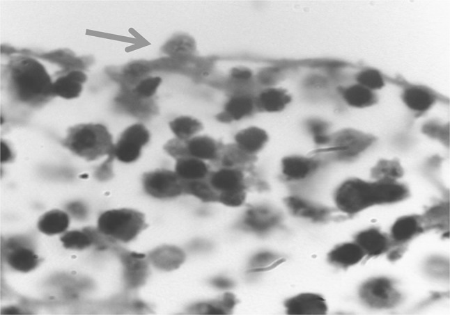
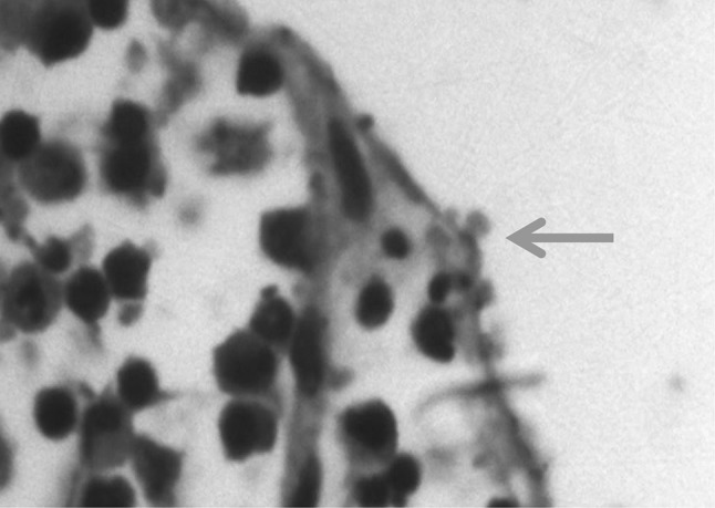
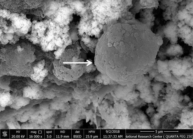
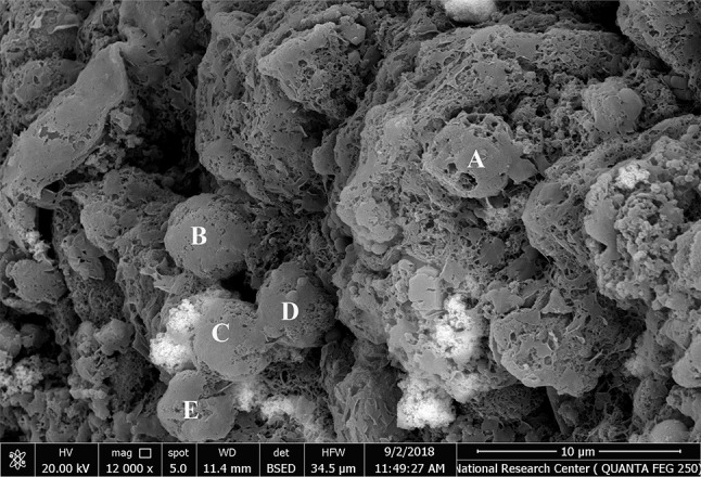
Cryptosporidium is an apicomplexan parasite of human and animals and is considered as an important co-factor in neonatal diarrhea. In this study, an explant culture was used as an in vitro model of buffalo intestine to evaluate the effect of Moringa leaves extract on Cryptosporidium parvum (C. parvum) oocysts using light and scanning electron microscopy and measuring IFN-γ, IL-12 and IL-14 in the culture supernatants. C. parvum oocysts were collected from naturally-infected calf feces, isolated, excysted and then co-inoculated with ileal tissue explants culture medium. The prepared Moringa leaves extract was then introduced to the infected tissues in the concentrations of 100 mg/ml and 300 mg/ml. After 24 h, tissues were collected and processed for light and scanning electron microscopy. Also, culture supernatants were collected for cytokines measurement. C. parvum parasitophorous vacuoles were found attached to the surface of tissue in Cryptosporidium-infected ileal tissue explants. High magnification imaging of ileal tissue explants using scanning electron microscopy showed that Moringa leaves extracts had a great effect on Cryptosporidium-infected ileal tissue explants. There was a high significant (P < 0.001) increase in IFN-γ, IL-12 and IL-14 (375, 275 and 90 pg/ml, respectively) in the supernatants of infected non-treated ileal tissue explant culture plate wells compared to the control non-infected ones (74.66, 75 and 50 pg/ml, respectively). A concentration of 100 mg/ml Moringa extract exhibited the highest anticryptosporidial effect causing a significant decrease in IFN-γ, IL-12 and IL-14 levels (225, 150 and 65 pg/ml, respectively) compared with supernatants of infected non-treated ileal explant culture plate wells. In this study, explant culturing of buffalo ileal tissues allowed investigating the pathogenesis of cryptosporidiosis using light and scanning electron microscopy and studying changes in cytokine levels in tissues with and without Moringa leaves extract treatment. This model could help to understand the regulation of intestinal secretory and inflammatory responses, and could be useful for the screening of potential anticryptosporidial candidate compounds.
Keywords: Buffaloes; Cryptosporidium parvum; IFN-γ; IL-12; IL-14; Moringa; Scanning electron microscopy; Tissue explant.
Conflict of interest statement
Conflict of interestThe authors declare that they have no conflicts or competing interests.
Figures


References
-
- Bauri RK, Tigga MN, Kullu SS. A review on use of medicinal plants to control parasites. Ind J Nat Prod Resour. 2015;6(4):268–277.
LinkOut - more resources
Full Text Sources