. 2012:2012:341629.
doi: 10.1155/2012/341629.

Analysis of NF-κB Pathway Proteins in Pediatric Hodgkin Lymphoma: Correlations with EBV Status and Clinical Outcome-A Children's Oncology Group Study

Affiliations

Analysis of NF-κB Pathway Proteins in Pediatric Hodgkin Lymphoma: Correlations with EBV Status and Clinical Outcome-A Children's Oncology Group Study

Terzah M Horton et al. Lymphoma. 2012.

Abstract

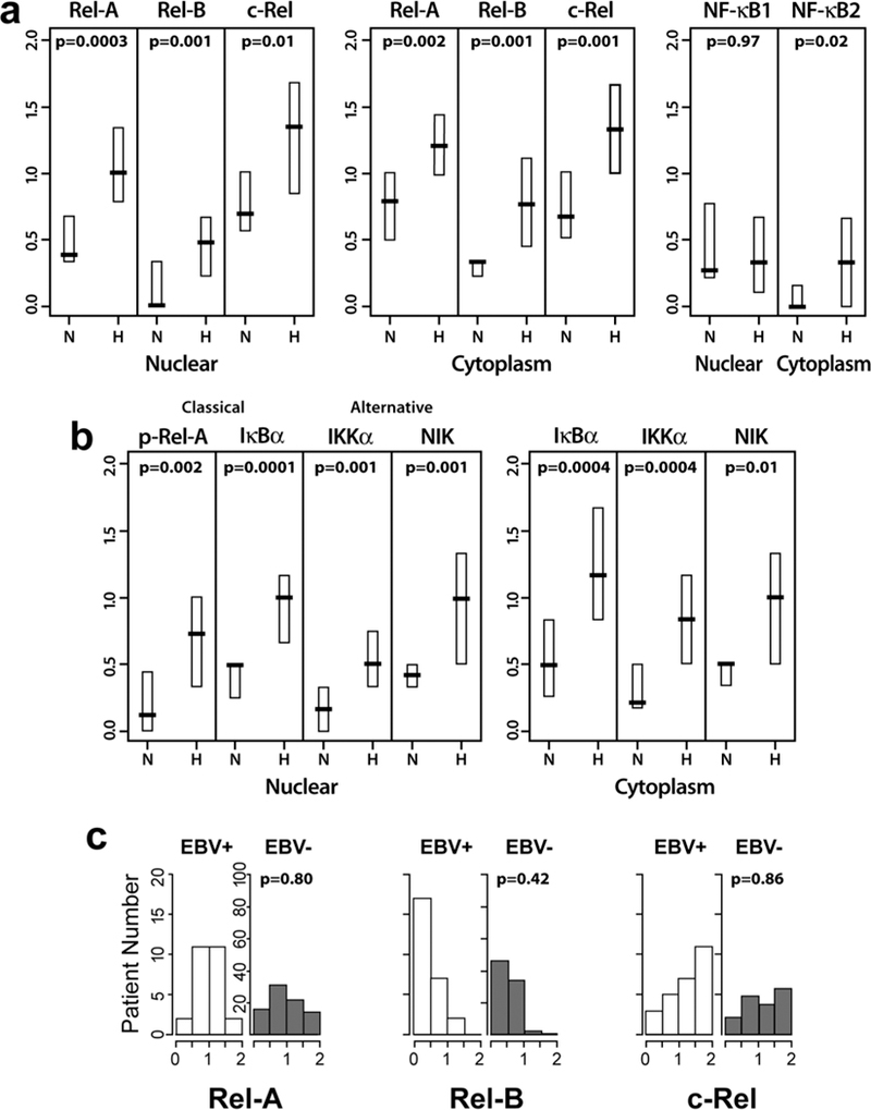
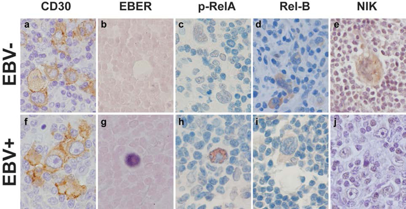
Constitutively active nuclear factor-κB (NF-κB) is integral to the survival of Hodgkin/Reed-Sternberg cells (H/RS) in Hodgkin Lymphoma (HL). To investigate NF-κB pathway proteins in pediatric HL, we utilized a tissue microarray compiled from 102 children enrolled in the Children's Oncology Group intermediate-risk clinical trial AHOD0031 (56 male, 78 Caucasian, median age 15y (range 1-20y), 85 nodular sclerosing subtype, 23 Epstein Barr virus (EBV) positive, 24 refractory/relapsed disease). We examined the intensity, localization, and pathway correlations of NF-κB pathway proteins (Rel-A/p65, Rel-B, c-Rel, NF-κB1, NF-κB2, IκB-α, IKK-α, IKK-β, IKK-γ/NEMO, NIK, A20), as well as their associations with EBV status and clinical outcome. NF-κB pathway proteins were overexpressed in pediatric HL patients compared to controls. Patients with EBV-tumors, or with rapid early therapy response, had tightly coordinated regulation of NF-κB pathway proteins, whereas patients with EBV+ tumors, or slow early therapy response, had little coordinated NF-κB pathway regulation. High NIK expression was associated with a slow response to therapy and decreased EFS. Elevated Rel-B, NIK and the NF-κB inhibitor A20 were associated with decreased EFS in multivariate analysis. These studies suggest a pivotal role for the NF-κB pathway in therapy response and patient survival (clinicaltrials.gov identifier: ).

Keywords: ABVD; AHOD0031; Epstein-Barr virus; Hodgkin Disease; NF-kappaB.


Conflict of interest statement

DISCLOSURES The authors report no potential conflicts of interest and no competing interests.

Figures

Figure 1:
Figure 1:. Classical and alternative NF-κB pathway activation
(A) Activation of the classical NF-κB pathway. Several receptors, including CD30, CD40 and LMP1, induce the classical NF-κB pathway by activating the IKK complex. Phosphorylated IKK-β phosphorylates IκB-α, inducing IκB polyubiquitination and degradation by the 26S proteasome. Following IκB degradation, NF-κB dimerizes with either c-Rel or NF-κB1/p50 and translocates into the nucleus where gene transcription is activated. (B) Activation of the alternative NF-κB pathway. The alternative NF-κB pathway is activated by a restricted set of cell-surface receptors, including CD30, CD40, and the BAFF receptor. Alternative NF-κB pathway activation increases the stability of NIK, which phosphorylates and activates IKK-α. IKK-α directly phosphorylates NF-κB2/p100, inducing partial proteolysis of p100 to p52 by the 26S proteasome. The p52 NF-κB subunit dimerizes with Rel-B and translocates into the nucleus, activating gene transcription. Proteins outlined in black were examined in the TMA. Cylindrical structure represents the 26S proteasome.
Figure 2:
Figure 2:. Comparison of NF-κB pathway protein intensities in HL patients with non-neoplastic controls.
(A) Nuclear and cytoplasmic NF-κB subunits from 102 HL samples (H) were compared to 10 controls (N). Significance was determined using the Wilcoxon rank-sum test. (B) Nuclear and cytoplasmic NF-κB pathway subunits phospho-Rel-A (p-Rel-A), IκB-α, IKKα, and NIK in HL samples compared to controls. (C) Intensity of nuclear NF-κB subunit expression in EBV+ tumors (white bars) and EBV-tumors (grey bars) for Rel-A, Rel-B and c-Rel. Patient average intensity scores (AIS) were placed in one of four protein intensity bins: 0–0.5 (very weak intensity), 0.5–1 (weak intensity), 1–1.5 (strong intensity) and 1.5–2 (very strong intensity).
Figure 3:
Figure 3:. Immunohistochemistry (IHC) and in situ hybridization in EBV+ vs. EBV-tumors.
CD30 (far left panel), EBER in situ hybridization, phospho (P)-Rel-A (center), Rel-B, and NIK (far right panel) in an EBV-patient (A) and an EBV+ patient (B). Pictures were taken at 400x magnification using an Olympus BX41 microscope. Whole images were corrected for contrast, brightness and color balance. Details of staining are provided in Supplemental Table I.
Figure 4:
Figure 4:. Spearman correlation coefficients between NF-κB pathway proteins stratified by EBV status (A) and rapidity of response to therapy (B).
Nuclear (a, b) and cytoplasmic (c, d) NF-κB pathway protein correlations were examined in EBV-patients (left panels) and EBV+ patients (right panels). Significant results are shown in white with raw p values, p value after correction for multiple comparisons are noted with asterisks (*p<0.05, **p<0.01). Full data set with Spearman correlation coefficients are shown in Supplemental Figure 3. (B) Nuclear (a, b) and cytoplasmic (c, d) NF-κB pathway protein correlations were examined in patients with rapid early response (RER) to ABVE-PC (left panels) vs. those with a slow early response (SER) the same therapy (right panels). Full data sets with Spearman correlation coefficients are shown in Supplemental Figure 4.
Figure 5:
Figure 5:. Examination of alternative NF-κB pathway proteins in HL patients with a slow early response (SER) to therapy.
(A) NIK expression in SER vs. RER: Left panel: Mean +/− 2 standard deviations (95% confidence intervals) of NIK levels in RER vs. SER. Significance was determined by the Wilcoxon rank-sum test. Middle and right panels: IHC from representative patients comparing NIK expression in an RER patient (middle panel) to an SER patient (right panel). (B) Alternative NF-κB pathway in SER samples: Scatter plot and linear regression analysis for cytoplasmic NIK (y-axis) vs. cytoplasmic IKK-α (left panel), cytoplasmic NF-κB2 (middle left panel), cytoplasmic Rel-B (middle right panel), and nuclear Rel-B (right panel). (C) Representative IHC from a SER patient samples immunostained with IKK-α (left panel), NF-κB2 (middle panel), and Rel-B (right panel). IHC pictures taken at 400x magnification using an Olympus BX41 microscope. Whole images were adjusted for contrast and brightness.

References

    1. Schmitz R, Stanelle J, Hansmann ML, Kuppers R. Pathogenesis of classical and lymphocyte-predominant Hodgkin lymphoma. Annu Rev Pathol 2009;4:151–74. - PubMed
    1. Kuppers R The biology of Hodgkin’s lymphoma. Nat Rev Cancer 2009. January;9(1):15–27. - PubMed
    1. Sun SC, Ley SC. New insights into NF-kappaB regulation and function. Trends Immunol 2008. October;29(10):469–78. - PMC - PubMed
    1. Mathas S, Lietz A, Anagnostopoulos I, Hummel F, Wiesner B, Janz M et al. c-FLIP mediates resistance of Hodgkin/Reed-Sternberg cells to death receptor-induced apoptosis. Journal of Experimental Medicine 2004;199(8):1041–52. - PMC - PubMed
    1. Kashkar H, Haefs C, Shin H, Hamilton-Dutoit SJ, Salvesen GS, Kronke M et al. XIAP-mediated caspase inhibition in Hodgkin’s lymphoma-derived B cells. J Exp Med 2003. July 21;198(2):341–7. - PMC - PubMed

LinkOut - more resources