Review
. 2019 Aug;38(4):389-408.
doi: 10.1007/s10930-019-09862-3.

The Rich Tapestry of Bacterial Protein Translocation Systems

Affiliations
Review

The Rich Tapestry of Bacterial Protein Translocation Systems

Peter J Christie. Protein J. 2019 Aug.

Abstract

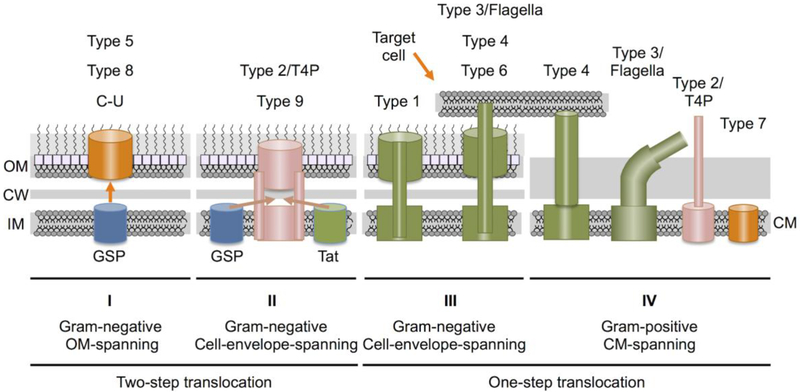
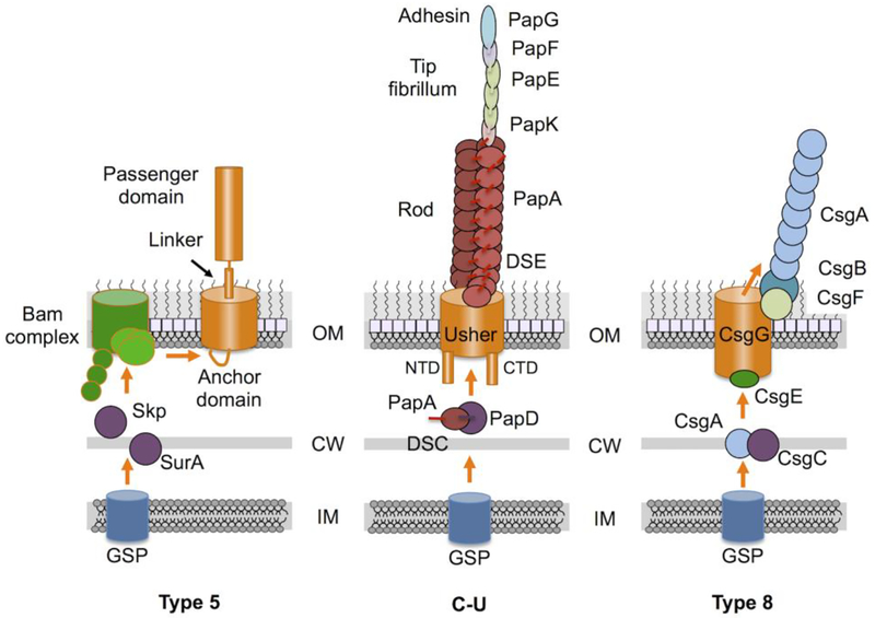
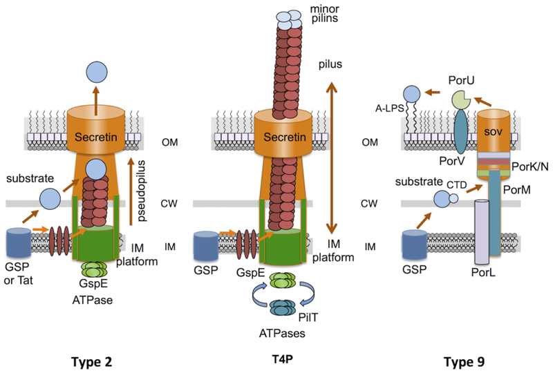
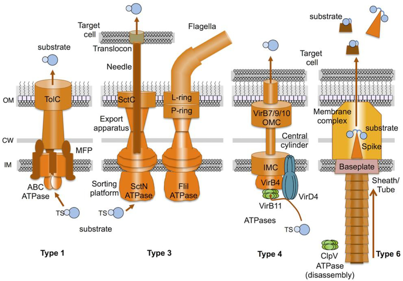
The translocation of proteins across membranes is a fundamental cellular function. Bacteria have evolved a striking array of pathways for delivering proteins into or across cytoplasmic membranes and, when present, outer membranes. Translocated proteins can form part of the membrane landscape, reside in the periplasmic space situated between the inner and outer membranes of Gram-negative bacteria, deposit on the cell surface, or be released to the extracellular milieu or injected directly into target cells. One protein translocation system, the general secretory pathway, is conserved in all domains of life. A second, the twin-arginine translocation pathway, is also phylogenetically distributed among most bacteria and plant chloroplasts. While all cell types have evolved additional systems dedicated to the translocation of protein cargoes, the number of such systems in bacteria is now known to exceed nine. These dedicated protein translocation systems, which include the types 1 through 9 secretion systems (T1SSs-T9SSs), the chaperone-usher pathway, and type IV pilus system, are the subject of this review. Most of these systems were originally identified and have been extensively characterized in Gram-negative or diderm (two-membrane) species. It is now known that several of these systems also have been adapted to function in Gram-positive or monoderm (single-membrane) species, and at least one pathway is found only in monoderms. This review briefly summarizes the distinctive mechanistic and structural features of each dedicated pathway, as well as the shared properties, that together account for the broad biological diversity of protein translocation in bacteria.

Keywords: Pathogenesis; Pilus; Protein translocation; Traffic ATPases.


Figures

Fig. 1.
Fig. 1.. Bacteria have evolved more than nine distinct pathways for delivering proteins across their cell envelopes.
Systems are grouped into one of four different Classes based on their location within the cell envelope and mechanism of transfer. Class I: These systems assemble at the outer membrane (OM) of Gram-negative bacteria and mediate substrate transfer across the OM. Class I systems fall into the broad category of two-step translocation systems because substrates are delivered to the cell surface in two-steps, first across the inner membrane (IM) via the general secretory pathway (GSP) and second across the outer membrane (OM) via the Class I machine. Class II: These systems assemble across the entire Gram-negative cell envelope but mediate substrate transfer only across the OM. Class II systems are also designated as two-step translocation systems because substrates are delivered across the IM via the GSP or twin-arginine-translocation (Tat) pathways, and across the OM by the Class II system. Class III: These systems assemble across the entire Gram-negative cell envelope and function as one-step translocation systems by recruiting substrates from the cytoplasm and delivering the cargoes to the cell surface through channels that span the IM, periplasm and OM. A subset of Class III machines deliver substrates directly into bacterial or eukaryotic target cells. Class IV: These systems assemble across the cytoplasmic membranes (CM) of Gram-positive bacteria. They generally function as one-step translocation systems by delivering substrates across the CM, although specialized features are also postulated to be necessary for translocation across the thick cell wall, or Mycobacterial spp. mycolic acid layer. Systems assigned to each Class are listed at top. C-U, chaperone-usher pathway; T4P, type 4 pilus assembly pathway.
Fig. 2.
Fig. 2.. Schematics of Class I assembly pathways.
Type 5 secretion systems (autotransporters) rely on the GSP for delivery across the IM and then are recruited to the Bam complex for OM insertion and surface display. C-U (Chaperone - Usher) systems as exemplified by the Pap pilus assembly pathway. Pilin subunits bind a periplasmic chaperone, e.g., PapD, by donor-strand complementation (DSC) and are delivered to the OM usher for delivery across the OM and pilus assembly by donor-strand exchange (DSE). Type 8 secretion systems are responsible for assembly of curli amyloid fibers.
Fig. 3.
Fig. 3.. Schematics of Class II assembly pathways.
Type 2 secretion systems elaborate ‘pseudopili’ on an IM platform. They recruit substrates delivered across the IM via the GSP or Tat systems to the tip of the pseudopilus, which might act as a piston to export the substrate through the secretin channel to the cell surface. T4Ps are phylogenetically and functionally related to the T2SSs, except they build pili that dynamically extend and retract through the coordinated actions of the GspE and PilT ATPases. Type 9 secretion systems recruit substrates from the periplasm through binding of conserved C-terminal domains (CTDs). Some substrates are covalently bound to the cell surface via a ‘sortase’ like mechanism.
Fig. 4.
Fig. 4.. Schematics of Class III assembly pathways.
Type 1 secretion systems are related to the ATP-binding cassette (ABC) transporter superfamily. They recruit substrates carrying C-terminal translocation signals (TSs) to the ATPase/MFP complex, and deliver substrates to the cell surface through the TolC channel/tunnel or ‘chunnel’. Type 3 systems consist of ‘injectisomes’ and phylogenetically-related flagella. Injectisomes elaborate injection needles for delivery of substrates into mammalian cell targets or pili for delivery to plant cell targets. Flagellar systems elaborate dynamic flagella for cellular chemotaxis. Substrates of injectisomes are recruited by recognition of N-terminal TSs or 5’ RNA structures with or without the aid of a chaperone. The SctN ATPase mediates unfolding of substrates prior to translocation through the needle. Type 4 systems are related to DNA conjugation machines. They elaborate cell envelope-spanning channels and deliver DNA or protein substrates via a contact-dependent mechanism into bacterial or eukaryotic target cells. Substrates are recognized by one or more TSs located at the C terminus or internally, with or without the aid of chaperones or adaptor proteins. Three ATPases (VirD4, VirB4, VirB11) are required for substrate translocation in most Gram-negative systems. Type 6 systems are bacteriophage contractile tail-like structures that are inverted for extrusion of substrates. Substrates associate covalently or noncovalently with the spike complex or the Hcp tube. The ClpV ATPase disassembles the sheath/tube complex after contraction and substrate ejection.

References

    1. Green ER, Mecsas J (2016) Bacterial secretion systems: An overview. Microbiol Spectr 4. doi: 10.1128/microbiolspec.VMBF-0012-2015. - DOI - PMC - PubMed
    1. Gerlach RG, Hensel M (2007) Protein secretion systems and adhesins: the molecular armory of Gram-negative pathogens. Int J Med Microbiol 297: 401–415. - PubMed
    1. Berks BC (2015) The twin-arginine protein translocation pathway. Annu Rev Biochem 84: 843–864. - PubMed
    1. Tsirigotaki A, De Geyter J, Sostaric N, Economou A, Karamanou S (2017) Protein export through the bacterial Sec pathway. Nat Rev Microbiol 15: 21–36. - PubMed
    1. Costa TR, Felisberto-Rodrigues C, Meir A, Prevost MS, Redzej A, et al. (2015) Secretion systems in Gram-negative bacteria: structural and mechanistic insights. Nat Rev Microbiol 13: 343–359. - PubMed

Publication types

MeSH terms

LinkOut - more resources