. 2019 Aug 13:8:e40855.
doi: 10.7554/eLife.40855.

Region-specific myelin differences define behavioral consequences of chronic social defeat stress in mice

Affiliations

Region-specific myelin differences define behavioral consequences of chronic social defeat stress in mice

Valentina Bonnefil et al. Elife. .

Abstract

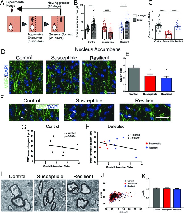
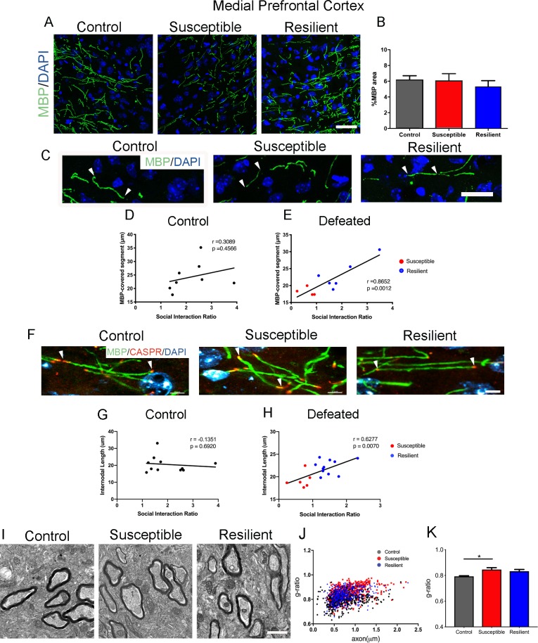
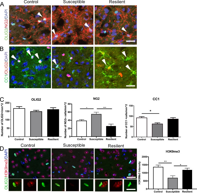
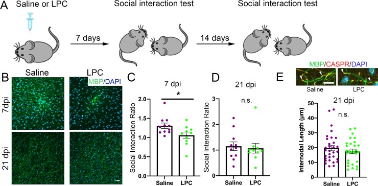
Exposure to stress increases the risk of developing mood disorders. While a subset of individuals displays vulnerability to stress, others remain resilient, but the molecular basis for these behavioral differences is not well understood. Using a model of chronic social defeat stress, we identified region-specific differences in myelination between mice that displayed social avoidance behavior ('susceptible') and those who escaped the deleterious effect to stress ('resilient'). Myelin protein content in the nucleus accumbens was reduced in all mice exposed to stress, whereas decreased myelin thickness and internodal length were detected only in the medial prefrontal cortex (mPFC) of susceptible mice, with fewer mature oligodendrocytes and decreased heterochromatic histone marks. Focal demyelination in the mPFC was sufficient to decrease social preference, which was restored following new myelin formation. Together these data highlight the functional role of mPFC myelination as critical determinant of the avoidance response to traumatic social experiences.

Editorial note: This article has been through an editorial process in which the authors decide how to respond to the issues raised during peer review. The Reviewing Editor's assessment is that all the issues have been addressed (see decision letter).

Keywords: epigenetics; medial prefrontal cortex; mouse; myelin; neuroscience; oligodendroccyte; resilience; social stress.


Conflict of interest statement

VB, KD, MA, MW, AA, JD, GT, HP, NB, PC, JL No competing interests declared

Figures

Figure 1.
Figure 1.. Effect of aggressive social encounters on myelination in the nucleus accumbens (NAc) of mice which showed two behaviorally distinct phenotypes following chronic social defeat stress (CSDS).
(A) The experimental paradigm for CSDS. (B–C) Mice susceptible to CSDS spent less time interacting with a conspecific mouse than the control group or resilient mice, as shown in (B) total time spent in the interaction zone when there is a conspecific mouse present and in (C) social interaction ratio defined by time spent in the interaction zone when a conspecific mouse present divided by a conspecific mouse absent. Control, n = 52; susceptible, n = 39; resilient, n = 33; ****, p<0.0001 by one-way ANOVA followed by Tukey’s post hoc test. (D–E) Representative confocal images and quantifications showing immunohistochemistry of myelin basic protein (MBP) counterstained with DAPI. Scale bar = 28 μm. n = 3 mice per group, two 20x images per animal; susceptible vs. control, p=0.0447; resilient vs. control, p=0.0109 by one-way ANOVA followed by Tukey’s post hoc test. (F) Representative confocal images showing MBP-covered myelinated segments. Arrowheads point to one MBP-covered myelinated segment. Scale car = 19 μm. (G–H) Pearson correlation coefficients showed non-significant correlation of MBP-covered segment length and social interaction ratio in control (G) or defeated (H) mice, control, 8 x-y pairs. r = −0.2242, p=0.5932. defeated, 10 x-y pairs, r = −0.3483, p=0.3240. control, n = 8 mice, susceptible, n = 4 mice, resilient, n = 6 mice, 1–2 20x images per mouse; (I) Representative electron microscopy images (scale bar = 1 μm) and (J–K) scatter plot and quantification of g-ratio; control, n = 5 mice; susceptible, n = 7 mice; resilient, n = 5 mice.
Figure 2.
Figure 2.. Myelination in the medial prefrontal cortex (mPFC) distinguished resilient from susceptible mice following stress.
(A–B) Representative confocal images and quantifications of myelin basic protein (MBP) counterstained with DAPI. Scale bar = 30 μm. n = 3 mice per group. Four 20x images taken per mouse (C) Representative confocal images showing MBP-covered myelinated segments. Arrowheads point to one continuous MBP-covered myelinated segment. Scale bar = 17 μm. (D–E) Pearson correlation coefficients showed significant correlation of MBP-covered segment length with social interaction ratio only in defeated (E) mice, but not in control (D), control, 8 x-y pairs, n = 8 mice, defeated 10 x-y pairs, susceptible, n = 4 mice, resilient, n = 6 mice, four 20x images were taken per mouse (F) Representative confocal images showing internodal segment marked by CASPR (Red) and MBP (Green). Arrowheads point to one internode. Scale bar = 5 μm. (G–H) Pearson correlation coefficients showed significant correlation of internodal length with social interaction ratio only in defeated (H) mice, but not in control (G), control, 11 x-y pairs, n = 11 mice, defeated 17 x-y pairs, susceptible, n = 6 mice, resilient, n = 11 mice, four-six 63x images taken per mouse. (I) Representative electron microscopy images, scale bar = 1 μm. (J–K) Scatter plot and quantification of g-ratio in the mPFC; control, n = 5 mice; susceptible, n = 7 mice; resilient, n = 5 mice; susceptible vs. control, p=0.0264 by one-way ANOVA followed by Tukey’s post hoc test.
Figure 3.
Figure 3.. Impaired oligodendrocyte differentiation was associated with reduced repressive histone methylation marks in the mPFC of susceptible mice.
(A–B) Representative confocal images of cells positive for OLIG2, NG2, and CC1 in the mPFC. DAPI was used as counterstain of nuclei. Scale bar = 25 μm. (C) quantification of OLIG2+ (n = 3 mice per group and 3–4 20x images taken per mouse), NG2+ cells (n = 3 mice per group and four images taken per mouse, *p=0.019, ***p<0.0001 by one-way ANOVA followed by Tukey’s post hoc test) and CC1+ cells (control, n = 8 mice, susceptible, n = 3 mice, resilient n = 7 mice, 3–4 20x images taken per mouse; *p=0.0195 by one-way ANOVA followed by Tukey’s post hoc test). (D) Representative confocal images and quantifications (E) of mean intensity of repressive histone mark H3K9me3 (Red) in OLIG2+ (Green) cells. control, n = 3 mice, susceptible, n = 2 mice, resilient, n = 5 mice, 4 20x images taken per mouse, 50–100 OLIG2+ cells were counted per image **p=0.0038, *p=0.0234 by one-way ANOVA followed by Tukey’s post hoc test. Data are mean ± S.E.M. Scale bar = 20 μm.
Figure 4.
Figure 4.. Focal demyelination in the mPFC reduced social preference behavior.
(A) The experimental paradigm for lysolecithin (LPC) injection and behavioral testing. (B) Representative confocal images showing reduced MBP immunointensity at seven dpi followed by a spontaneous restoration at 21 dpi. (C) Mice received LPC displayed reduced social preference behavior at seven dpi as quantified by social interaction ratio. (D) Restoration of normal social interaction behavior at 21 dpi as quantified by social interaction ratio. Saline, n = 11 mice; LPC, n = 10 mice *, p<0.05 by unpaired t-test. Data are mean ± S.E.M. (E) Representative confocal images of immunohistochemistry of MBP (Green) and CASPR (Red) and scatter plots of internodal length at 21dpi. Counterstained with DAPI. Saline n = 3 mice, LPC, n = 5 mice. 2–4 63x images taken per mouse.

References

    1. Berton O, McClung CA, Dileone RJ, Krishnan V, Renthal W, Russo SJ, Graham D, Tsankova NM, Bolanos CA, Rios M, Monteggia LM, Self DW, Nestler EJ. Essential role of BDNF in the mesolimbic dopamine pathway in social defeat stress. Science. 2006;311:864–868. doi: 10.1126/science.1120972. - DOI - PubMed
    1. Cathomas F, Azzinnari D, Bergamini G, Sigrist H, Buerge M, Hoop V, Wicki B, Goetze L, Soares S, Kukelova D, Seifritz E, Goebbels S, Nave KA, Ghandour MS, Seoighe C, Hildebrandt T, Leparc G, Klein H, Stupka E, Hengerer B, Pryce CR. Oligodendrocyte gene expression is reduced by and influences effects of chronic social stress in mice. Genes, Brain and Behavior. 2019;18:e12475. doi: 10.1111/gbb.12475. - DOI - PubMed
    1. Covington HE, Lobo MK, Maze I, Vialou V, Hyman JM, Zaman S, LaPlant Q, Mouzon E, Ghose S, Tamminga CA, Neve RL, Deisseroth K, Nestler EJ. Antidepressant effect of optogenetic stimulation of the medial prefrontal cortex. Journal of Neuroscience. 2010;30:16082–16090. doi: 10.1523/JNEUROSCI.1731-10.2010. - DOI - PMC - PubMed
    1. Dowlati Y, Herrmann N, Swardfager W, Liu H, Sham L, Reim EK, Lanctôt KL. A meta-analysis of cytokines in major depression. Biological Psychiatry. 2010;67:446–457. doi: 10.1016/j.biopsych.2009.09.033. - DOI - PubMed
    1. Erta M, Quintana A, Hidalgo J. Interleukin-6, a major cytokine in the central nervous system. International Journal of Biological Sciences. 2012;8:1254–1266. doi: 10.7150/ijbs.4679. - DOI - PMC - PubMed

Publication types

Substances