. 2020 Feb;287(3):452-464.
doi: 10.1111/febs.15040. Epub 2019 Aug 28.

Proteolytic and nonproteolytic activation mechanisms result in conformationally and functionally different forms of coagulation factor XIII A

Affiliations

Proteolytic and nonproteolytic activation mechanisms result in conformationally and functionally different forms of coagulation factor XIII A

Boris A Anokhin et al. FEBS J. 2020 Feb.

Abstract

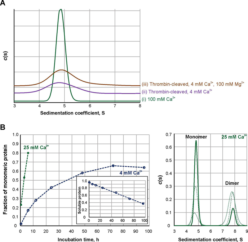
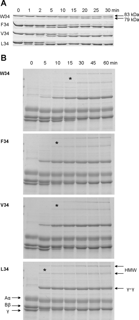
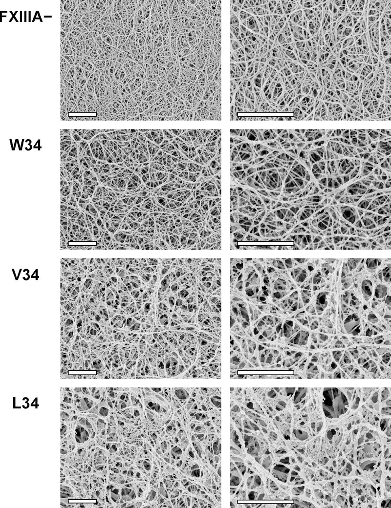
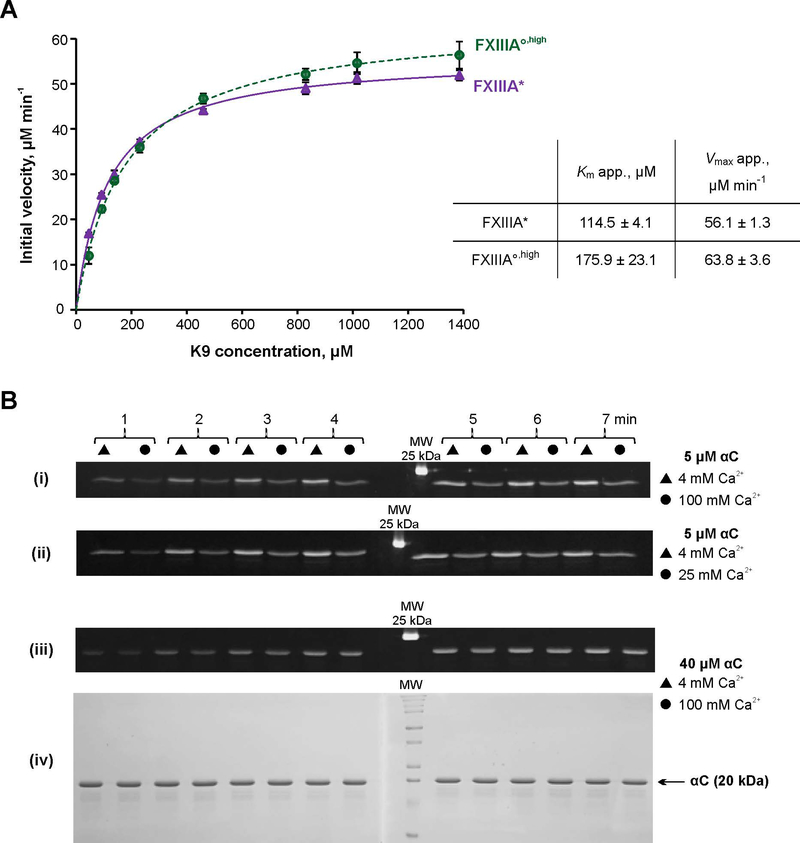
Factor XIIIA (FXIIIA) is a transglutaminase that cross-links intra- and extracellular protein substrates. FXIIIA is expressed as an inactive zymogen, and during blood coagulation, it is activated by removal of an activation peptide by the protease thrombin. No such proteolytic FXIIIA activation is known to occur in other tissues or the intracellular form of FXIIIA. For those locations, FXIIIA is assumed instead to undergo activation by Ca2+ ions. Previously, we demonstrated a monomeric state for active FXIIIA. Current analytical ultracentrifugation and kinetic experiments revealed that thrombin-activated FXIIIA has a higher conformational flexibility and a stronger affinity toward glutamine substrate than does nonproteolytically activated FXIIIA. The proteolytic activation of FXIIIA was further investigated in a context of fibrin clotting. In a series of fibrin cross-linking assays and scanning electron microscopy studies of plasma clots, the activation rates of FXIIIA V34X variants were correlated with the extent of fibrin cross-linking and incorporation of nonfibrous protein into the clot. Overall, the results suggest conformational and functional differences between active FXIIIA forms, thus expanding the understanding of FXIIIA function. Those differences may serve as a basis for developing therapeutic strategies to target FXIIIA in different physiological environments. ENZYMES: Factor XIIIA ( EC 2.3.2.13).

Keywords: analytical ultracentrifugation; factor XIII; fibrin clot; scanning electron microscopy; transglutaminase.


Conflict of interest statement

Conflict of interest The authors state that they have no conflict of interests.

Figures

Fig. 1
Fig. 1. Sedimentation properties and activation rate of FXIIIA under different conditions.
A – Sedimentation profiles of FXIIIA studied by AUC: 2 μM FXIIIA was activated at 37 °C for 30 min nonproteolytically by 100 mM CaCl2 (trace I, green) or proteolytically by 3.5 NIH units/ml bovine thrombin in the presence of 4 mM CaCl2 (trace ii, purple). An additional sample of thrombin-activated FXIIIA contained 4 mM CaCl2 and 100 mM MgCl2 (trace iii, orange). Two independent samples were analyzed for each condition, with the same results. (N=2) B – Dissociation progress of 2 μM FXIIIA in the presence of 4 (open blue circles) and 25 mM CaCl2 (filled green circles). Graph on the left presents quantitative analysis of sedimentation velocity AUC data. The insert demonstrates fraction of soluble protein (estimated from absorbance at 280 nm) in samples of 4 mM Ca2+-activated FXIIIA as a function of time. The panel on the right depicts AUC sedimentation profiles for the FXIIIA samples incubated in the presence of 25 mM CaCl2 for 30 min (dotted green line), 3 h (dashed green line) and 6 h (solid green line).
Fig. 2
Fig. 2. Effect of the rate of FXIIIA proteolytic activation on fibrin crosslinking.
A – 1 μM recombinant FXIIIA AP variants were incubated with 30 nM recombinant human thrombin at 37 °C. Aliquots were withdrawn at denoted time points and thrombin was inhibited with 760 nM PPACK. The samples were then subjected to SDS-PAGE on 8% gels. FXIIIA AP cleavage resulting in a 4 kDa molecular weight loss could then be followed by monitoring the appearance of a 79 kDa band. (N=3) B – Fibrin crosslinking by FXIIIA AP variants. 1 mg/ml human FXIII-free fibrinogen was combined with a recombinant FXIIIA AP-variant (50 nM) at 37 °C, and crosslinking was initiated by addition of 12 nM recombinant human thrombin and 2.5 mM CaCl2. At each denoted time point, the reaction was stopped by addition of reducing sample loading buffer and boiling. The samples were resolved via SDS-PAGE (8% gel). Fibrinogen chains Aα, Bβ, γ, HMW (high molecular weight crosslinks), and γ–γ crosslinks are annotated. * symbol denotes the earliest detection of HMW species during the course of the crosslinking reaction. (N = 3)
Fig. 3
Fig. 3. Scanning electron microscopy of fibrin clots in the presence of FXIIIA AP variants.
100 nM FXIIIA AP variants were combined with plasma from FXIIIA-deficient mice (final dilution of plasma was 1:4). Control samples were made without FXIIIA (FXIIIA ). Clotting was initiated by addition of 2.1 NIH units/ml bovine thrombin and 13.5 mM CaCl2. Clots were formed for 2 h at 37 °C and prepared for SEM as described in Materials and Methods. For each FXIIIA AP variant, two clots were studied, with essentially the same results. Each set of clots (FXIII , W34, V34, and L34) was formed using plasma obtained from the same mouse. Shown are representative SEM photographs at two magnifications: 10,000x (left) and 20,000x (right), scale bars are 2 μm. Note that during the SEM sample preparation process, the fixative agent glutaraldehyde was not included to avoid artificial protein crosslinking. As a result, the FXIIIA clots were significantly flattened during the dehydration process. In response, the resultant FXIIIA fibrin fibers on the SEM images appeared denser packed.
Fig. 4
Fig. 4. Transglutaminase activity of FXIIIA under different conditions.
A – Spectrophotometric kinetic assay. Glutamine donor K9 peptide was preincubated with the lysine mimic DMPDA at 37 °C for 5 min, and the crosslinking reaction was initiated by addition of 1 μM FXIIIA°,high (green circle) or FXIIIA* (purple circle). The reaction was followed by an increase in absorbance at 278 nm due to formation of the anilide crosslinked product. The plot represents Michaelis-Menten fits of the initial reaction velocities as function of the K9 concentration. Apparent Km and Vmax resulting from the fits are shown in the table insert. Values are presented as mean ± SD (N=3). B – MDC crosslinking SDS-PAGE based assay. Recombinant fibrinogen αC (233–425) was preincubated with 1 mM MDC at 37 °C for 5 min. The crosslinking reaction was initiated by addition of 100 nM FXIIIA°,high or FXIIIA*. Reaction aliquots were withdrawn at 1 – 7 min and quenched by addition of reducing sample buffer and boiling. The time point samples were loaded on 15% SDS-PAGE side by side for FXIIIA* (triangles) and FXIIIA°,high (circles). Two gels were run (1 – 4 and 5 – 7 min time points). The gels were aligned and photographed under UV light (panels i – iii). Panel iv – Coomassie Blue-stained gel pair from panel iii demonstrating absence of αC–αC conjugation. FXIIIA* was always preactivated in the presence of 4 mM CaCl2, and FXIIIA°,high was preactivated in the presence of 100 mM CaCl2. Concentrations of αC and CaCl2 in the crosslinking reaction mix are annotated on the right. The different MDC assay series were performed three independent times.

References

    1. Komaromi I, Bagoly Z & Muszbek L (2011) Factor XIII: novel structural and functional aspects, Journal of thrombosis and haemostasis : JTH. 9, 9–20. - PubMed
    1. Muszbek L, Bereczky Z, Bagoly Z, Komaromi I & Katona E (2011) Factor XIII: a coagulation factor with multiple plasmatic and cellular functions, Physiological reviews. 91, 931–72. - PubMed
    1. Schroeder V & Kohler HP (2016) Factor XIII: Structure and Function, Seminars in thrombosis and hemostasis. 42, 422–8. - PubMed
    1. Eckert RL, Kaartinen MT, Nurminskaya M, Belkin AM, Colak G, Johnson GV & Mehta K (2014) Transglutaminase regulation of cell function, Physiol Rev. 94, 383–417. - PMC - PubMed
    1. Hoac B, Nelea V, Jiang W, Kaartinen MT & McKee MD (2017) Mineralization-inhibiting effects of transglutaminase-crosslinked polymeric osteopontin, Bone. 101, 37–48. - PubMed

Publication types