. 2019 Sep 7;11(33):15647-15658.
doi: 10.1039/c9nr04828f. Epub 2019 Aug 13.

Contrast enhanced ultrasound imaging by nature-inspired ultrastable echogenic nanobubbles

Affiliations

Contrast enhanced ultrasound imaging by nature-inspired ultrastable echogenic nanobubbles

Al de Leon et al. Nanoscale. .

Abstract

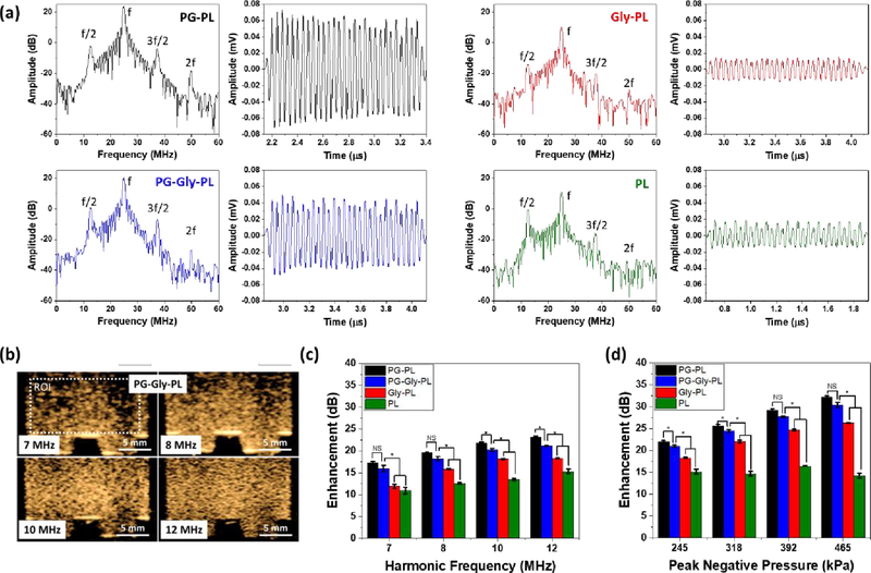
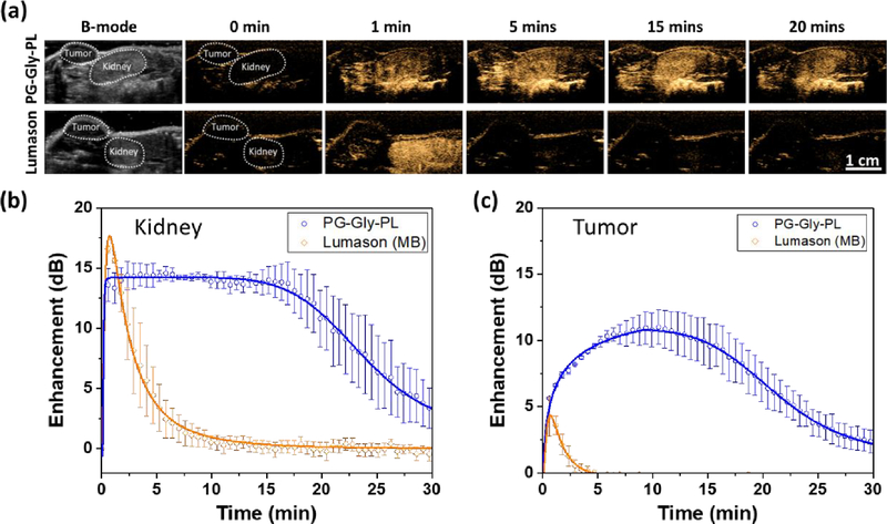
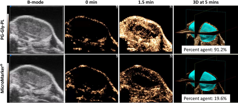
Advancement of ultrasound molecular imaging applications requires not only a reduction in size of the ultrasound contrast agents (UCAs) but also a significant improvement in the in vivo stability of the shell-stabilized gas bubble. The transition from first generation to second generation UCAs was marked by an advancement in stability as air was replaced by a hydrophobic gas, such as perfluoropropane and sulfur hexafluoride. Further improvement can be realized by focusing on how well the UCAs shell can retain the encapsulated gas under extreme mechanical deformations. Here we report the next generation of UCAs for which we engineered the shell structure to impart much better stability under repeated prolonged oscillation due to ultrasound, and large changes in shear and turbulence as it circulates within the body. By adapting an architecture with two layers of contrasting elastic properties similar to bacterial cell envelopes, our ultrastable nanobubbles (NBs) withstand continuous in vitro exposure to ultrasound with minimal signal decay and have a significant delay on the onset of in vivo signal decay in kidney, liver, and tumor. Development of ultrastable NBs can potentially expand the role of ultrasound in molecular imaging, theranostics, and drug delivery.


Conflict of interest statement

Conflict of Interest

The authors declare no conflict of interest.

Figures

Figure 1.
Figure 1.. Schematic, size, concentration, and structural property of NBs.
(a) Schematic representation of the membrane of the ultrastable NB showing a bilayer architecture with elastic contrast. (b). Cryo-electron microscopy (cryo-EM) images of PG-Gly-PL showing the nanobubble membrane and the dense C3F8 gas core. Diameter (c) and concentration (d) measurement of NB via a resonant mass measurement capable of differentiating buoyant (NBs) and non-buoyant particles (micelles, liposomes, etc.). Mean ± SE (n = 5) (e) X-ray diffractogram of the membrane of the freeze-dried NBs. The NBs were isolated and freeze-dried overnight. Peak corresponding to d = 4.5 Å is observed for all NB but only PG-PL and PG-Gly-PL have a peak corresponding to d = 10.3 Å implying the incorporation of PG into the phospholipid membrane.
Figure 2.
Figure 2.. Nonlinear oscillation of a single NB and a solution of NBs.
(a) Representative nonlinear backscatter from a single NB from a narrowband 30 cycle ultrasound pulse and the corresponding frequency spectrum (25 MHz transducer at 300kPa). Presence of subharmonic (f/2), ultraharmonic (3f/2), and second harmonic (2f), in addition to its fundamental (f) harmonic signal in the frequency spectra confirms the nonlinear oscillation of NBs. Minor changes in backscattered amplitude during the 30 cycle pulse shows how stably PG-Gly-PL NB oscillates when insonated by 25 MHz ultrasound. The magnitude of amplitude is consistent with the predicted stiffness of the NB shell having PG-PL as the most flexible, Gly-PL as the stiffest, and PG-Gly-PL with intermediate stiffness. (b) Contrast harmonic images (2nd harmonic) of PG-Gly-PL at different receiving frequency showing improvement in both intensity and resolution of the backscattered signal. NB with concentration of 4 × 10⁹ NBs/mL was placed in a tissue-mimicking agarose phantom. Only the second harmonic backscattered signal is visible in contrast harmonic imaging mode which implies that the backscattered signal from the agarose phantom, similar to tissues, does not have non-linear component. Enhancement of nonlinear backscattered signal of NB (concentration of 4 × 10⁹ NB/mL) relative to tissue-mimicking agarose phantom at different (c) receiving frequency and (d) peak negative pressure.
Figure 3.
Figure 3.. In vitro characterization of NBs in tissue mimicking agarose phantom.
(a) Contrast harmonic images of PG-Gly-PL (concentration of 4 × 10⁹ NB/mL) before and after application of 500 frames of ultrasound at different peak negative pressure (P = 245 kPa to 465 kPa) at 1 frame per second. A dilute solution of PG-Gly-PL NB is placed in a tissue-mimicking agarose phantom with a thin channel (1 mm) to ensure minimal diffusion of NB in and out of the ultrasound field. (b) Representative signal decay curve of NBs continuously exposed to 245 kPa pressure at 1 frame per second. Mean ± SE (n = 3). (c) In vitro half-life of nonlinear backscattered signal of dilute NBs placed in a tissue mimicking agarose phantom and at 245 kPa pressure and varying frame rate. Mean ± SE (n = 3). (d) In vitro half-life (t1/2) of nonlinear backscattered signal dilute NBs placed in a tissue mimicking agarose phantom and exposed to ultrasound with varying peak negative pressure at 15 frames per second. Mean ± SE (n = 3).
Figure 4.
Figure 4.. In vivo characterization of NBs in mouse kidney stability relative to commercially available UCA in mice
(a) NBs and commercially available UCA (Lumason) were injected via tail-vein and a cross-section of the kidney and liver was imaged at 12 MHz, 245 kPa pressure, and 0.2 frames per second. Left: Representative B-mode images of the kidney and liver before the injection of UCAs. 0 min-20 mins: Series of images showing the signal onset and signal decay of various UCAs at different time points. Minimal to no contrast can be observed before injection (t= 0) which is expected because backscatter signals from kidney and liver do not have any nonlinear properties. (b) Representative signal decay curve of NBs and Lumason® showing the delayed signal decay onset and longer in vivo half-life of PG-Gly-PL. (c) In vivo maximum enhancement, (d) in vivo washout half-life, and (e) in vivo decay onset of NBs and Lumason extracted from the enhancement vs time curve.
Figure 5.
Figure 5.. In vivo characterization of NBs in mouse kidney and flank colorectal tumor stability relative to commercially available UCA (Lumason®) in mice.
(a) NBs or Lumason® were injected via tail-vein and a cross-section of the kidney and flank tumor was imaged at 12 MHz, 245 kPa pressure, and 0.2 frames per second. Left: Representative B-mode images of the kidney and tumor before the injection of UCAs. 0 min-20 mins: Series of images showing the signal onset and signal decay of various UCAs at different time points. Minimal to no contrast can be observed before injection (t= 0) which is expected because backscatter signals from kidney and tumor do not have any nonlinear properties. Representative signal decay curves of PG-Gly-PL and Lumason® in kidney (b) and in tumor (c) showing the delayed signal decay onset and longer in vivo half-life of PG-Gly-PL.
Figure 6.
Figure 6.
In vivo characterization of PG-Gly-PL in flank colorectal tumor stability relative to MicroMarker® in mice showing comparison on the extent of UCA filling of tumor both in 2D and 3D. Tumor volume: 229.975 mm3.

References

    1. Szabo T, No TitleDiagnostic Ultrasound Imaging: Inside Out, Academic Press, Second Edi., 2013.
    1. Paproski RJ, Forbrich A, Huynh E, Chen J, Lewis JD, Zheng G and Zemp RJ, Small, 2016, 12, 371–380. - PubMed
    1. Wang X, Chen H, Zhang K, Ma M, Li F, Zeng D, Zheng S, Chen Y, Jiang L, Xu H and Shi J, Small, 2014, 10, 1403–1411. - PubMed
    1. Hadinger KP, Marshalek JP, Sheeran PS, Dayton PA and Matsunaga TO, Ultrasound Med. Biol, 2018, 44, 2728–2738. - PMC - PubMed
    1. Rojas JD and Dayton PA, Ultrasound Med. Biol, 2019, 45, 177–191. - PubMed