

Association of Intensive vs Standard Blood Pressure Control With Cerebral White Matter Lesions
- PMID: 31408137
- PMCID: PMC6692679
- DOI: 10.1001/jama.2019.10551
Association of Intensive vs Standard Blood Pressure Control With Cerebral White Matter Lesions
Abstract
Importance: The effect of intensive blood pressure lowering on brain health remains uncertain.
Objective: To evaluate the association of intensive blood pressure treatment with cerebral white matter lesion and brain volumes.
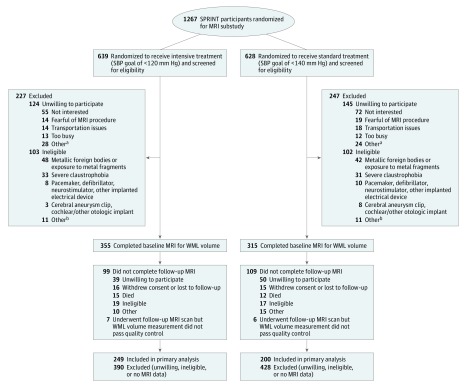
Design, setting, and participants: A substudy of a multicenter randomized clinical trial of hypertensive adults 50 years or older without a history of diabetes or stroke at 27 sites in the United States. Randomization began on November 8, 2010. The overall trial was stopped early because of benefit for its primary outcome (a composite of cardiovascular events) and all-cause mortality on August 20, 2015. Brain magnetic resonance imaging (MRI) was performed on a subset of participants at baseline (n = 670) and at 4 years of follow-up (n = 449); final follow-up date was July 1, 2016.
Interventions: Participants were randomized to a systolic blood pressure (SBP) goal of either less than 120 mm Hg (intensive treatment, n = 355) or less than 140 mm Hg (standard treatment, n = 315).
Main outcomes and measures: The primary outcome was change in total white matter lesion volume from baseline. Change in total brain volume was a secondary outcome.
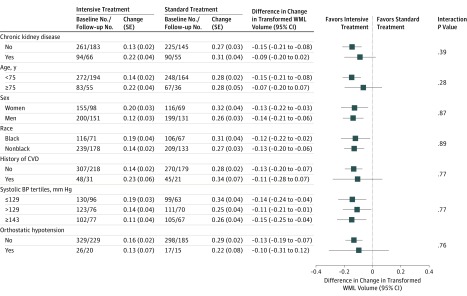
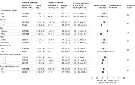
Results: Among 670 recruited patients who had baseline MRI (mean age, 67.3 [SD, 8.2] years; 40.4% women), 449 (67.0%) completed the follow-up MRI at a median of 3.97 years after randomization, after a median intervention period of 3.40 years. In the intensive treatment group, based on a robust linear mixed model, mean white matter lesion volume increased from 4.57 to 5.49 cm3 (difference, 0.92 cm3 [95% CI, 0.69 to 1.14]) vs an increase from 4.40 to 5.85 cm3 (difference, 1.45 cm3 [95% CI, 1.21 to 1.70]) in the standard treatment group (between-group difference in change, -0.54 cm3 [95% CI, -0.87 to -0.20]). Mean total brain volume decreased from 1134.5 to 1104.0 cm3 (difference, -30.6 cm3 [95% CI, -32.3 to -28.8]) in the intensive treatment group vs a decrease from 1134.0 to 1107.1 cm3 (difference, -26.9 cm3 [95% CI, 24.8 to 28.8]) in the standard treatment group (between-group difference in change, -3.7 cm3 [95% CI, -6.3 to -1.1]).
Conclusions and relevance: Among hypertensive adults, targeting an SBP of less than 120 mm Hg, compared with less than 140 mm Hg, was significantly associated with a smaller increase in cerebral white matter lesion volume and a greater decrease in total brain volume, although the differences were small.
Trial registration: ClinicalTrials.gov Identifier: NCT01206062.
Conflict of interest statement
Figures
Comment in
-
Blood Pressure, Brain Volume and White Matter Hyperintensities, and Dementia Risk.JAMA. 2019 Aug 13;322(6):512-513. doi: 10.1001/jama.2019.10849. JAMA. 2019. PMID: 31408120 No abstract available.
References
-
- Basile AM, Pantoni L, Pracucci G, et al. ; LADIS Study Group . Age, hypertension, and lacunar stroke are the major determinants of the severity of age-related white matter changes: the LADIS (Leukoaraiosis and Disability in the Elderly) study. Cerebrovasc Dis. 2006;21(5-6):315-322. doi: 10.1159/000091536 - DOI - PubMed
-
- Gorelick PB, Scuteri A, Black SE, et al. ; American Heart Association Stroke Council, Council on Epidemiology and Prevention, Council on Cardiovascular Nursing, Council on Cardiovascular Radiology and Intervention, and Council on Cardiovascular Surgery and Anesthesia . Vascular contributions to cognitive impairment and dementia: a statement for healthcare professionals from the American Heart Association/American Stroke Association. Stroke. 2011;42(9):2672-2713. doi: 10.1161/STR.0b013e3182299496 - DOI - PMC - PubMed
Publication types
MeSH terms
Substances
Associated data
Grants and funding
- UL1 TR000439/TR/NCATS NIH HHS/United States
- UL1 TR002548/TR/NCATS NIH HHS/United States
- UL1 TR000005/TR/NCATS NIH HHS/United States
- UL1 TR000073/TR/NCATS NIH HHS/United States
- UL1 RR024134/RR/NCRR NIH HHS/United States
- P30 AG049638/AG/NIA NIH HHS/United States
- UL1 TR000003/TR/NCATS NIH HHS/United States
- UL1 TR000105/TR/NCATS NIH HHS/United States
- RF1 AG054409/AG/NIA NIH HHS/United States
- UL1 TR002240/TR/NCATS NIH HHS/United States
- UL1 TR000093/TR/NCATS NIH HHS/United States
- UL1 TR000075/TR/NCATS NIH HHS/United States
- P30 GM103337/GM/NIGMS NIH HHS/United States
- UL1 TR000064/TR/NCATS NIH HHS/United States
- P41 RR013642/RR/NCRR NIH HHS/United States
- UL1 TR000050/TR/NCATS NIH HHS/United States
- UL1 TR002003/TR/NCATS NIH HHS/United States
- UL1 RR025755/RR/NCRR NIH HHS/United States
- UL1 TR000433/TR/NCATS NIH HHS/United States
- UL1 TR000002/TR/NCATS NIH HHS/United States
- UL1 TR001064/TR/NCATS NIH HHS/United States
- UL1 TR000445/TR/NCATS NIH HHS/United States
- UL1 TR003142/TR/NCATS NIH HHS/United States
- UL1 RR025771/RR/NCRR NIH HHS/United States
LinkOut - more resources
Full Text Sources
Other Literature Sources
Medical
