Review
. 2019 Dec;107(12):2736-2755.
doi: 10.1002/jbm.a.36777. Epub 2019 Aug 27.

Three-dimensional cryogels for biomedical applications

Affiliations
Review

Three-dimensional cryogels for biomedical applications

Mehdi Razavi et al. J Biomed Mater Res A. 2019 Dec.

Abstract

Cryogels are a subset of hydrogels synthesized under sub-zero temperatures: initially solvents undergo active freezing, which causes crystal formation, which is then followed by active melting to create interconnected supermacropores. Cryogels possess several attributes suited for their use as bioscaffolds, including physical resilience, bio-adaptability, and a macroporous architecture. Furthermore, their structure facilitates cellular migration, tissue-ingrowth, and diffusion of solutes, including nano- and micro-particle trafficking, into its supermacropores. Currently, subsets of cryogels made from both natural biopolymers such as gelatin, collagen, laminin, chitosan, silk fibroin, and agarose and/or synthetic biopolymers such as hydroxyethyl methacrylate, poly-vinyl alcohol, and poly(ethylene glycol) have been employed as 3D bioscaffolds. These cryogels have been used for different applications such as cartilage, bone, muscle, nerve, cardiovascular, and lung regeneration. Cryogels have also been used in wound healing, stem cell therapy, and diabetes cellular therapy. In this review, we summarize the synthesis protocol and properties of cryogels, evaluation techniques as well as current in vitro and in vivo cryogel applications. A discussion of the potential benefit of cryogels for future research and their application are also presented.

Keywords: bioscaffolds; cellular therapies; cryogels; tissue engineering.


Figures

FIGURE 1
FIGURE 1
Schematic representation of the cryogelation process: (a) a solution of monomers and/or polymers with or without the presence of a cross-linker; (b) freezing process; (c) cryogel wall synthesis occurs at the “unfrozen liquid microphase” at sub-zero temperatures; (d) thawing process; (e) formation of mature cryogel with macroporous network in native hydration state
FIGURE 2
FIGURE 2
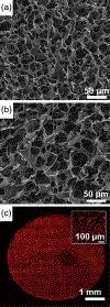
Microarchitecture of gelatin-based cryogels: (a) surface and (b) cross-sectional SEM micrograph images of highly-porous gelatin cryogels; (c) 2-photon imaging at a depth of 150 μm below the surface of a rhodamine-gelatin cryogel, with a magnified inset view at the center of the bioscaffold diameter (reproduced with permission from Koshy, S. T., Ferrante, T. C., Lewin, S. A., & Mooney, D. J. (2014). Injectable, porous, and cell-responsive gelatin cryogels. Biomaterials, 35, 2477–2487)
FIGURE 3
FIGURE 3
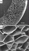
(a) SEM cross-sectional micrograph views of agarose–alginate cryogel sheets, and (b) a magnified image demonstrating the uniform pore size distribution present within the sheet format (reproduced with permission from Tripathi, A. & Kumar, A. (2011). Multi-featured macroporous Agarose-alginate cryogel: Synthesis and characterization for bioengineering applications. Macromolecular Bioscience, 11, 22–35)
FIGURE 4
FIGURE 4
(a) Photographs, μ-CT, SEM images of an uncoated- and polydopamine coated-cryogel bioscaffold: In μ-CT images, the purple areas show the bioscaffold material and the dark areas refer to the void space, SEM images showing the existence of both small, large, and continuously interconnected macropores throughout the entire bioscaffold construct; (b) SEM images of AD-MSCs (indicated by white arrows) on the superficial layer and within the center of uncoated- and polydopamine coated-cryogel bioscaffolds at Day 10; (c) confocal images of AD-MSCs after 1 and 10 days seeding into uncoated- and polydopamine coated-cryogel bioscaffolds (direct contact) and culturing with the medium of uncoated- and polydopamine coated-cryogel bioscaffolds (indirect contact) and the results of AD-MSCs counting at Day 0, 1, and 10 showing the improved AD-MSCs proliferation when cryogel bioscaffolds were coated with polydopamine (reproduced with permission from Razavi, M., Hu, S., & Thakor, A. S. (2018). A collagen based cryogel bioscaffold coated with nanostructured polydopamine as a platform for mesenchymal stem cell therapy. Journal of Biomedical Materials Research Part A, 106, 2213–2228). Significant differences: *p < .05, difference between the control group and bioscaffolds. #p < .05, difference between uncoated- and polydopamine coated-cryogel bioscaffolds. (Unpaired Student’s t-test)
FIGURE 5
FIGURE 5
(a) Schematic representation of different techniques for cryogel bioscaffold cell-seeding include static and perfusion (dynamic) methods (reproduced with permission from Petrenko, Y. A., Ivanov, R. V., Lozinsky, V. I., & Petrenko, A. Y. (2011). Comparison of the methods for seeding human bone marrow mesenchymal stem cells to macroporous alginate cryogel carriers. Bulletin of Experimental Biology and Medicine, 150, 543–546); (b) schematic of artificial cryogel bioscaffolding sliced into 100 μm sections placed into a cell-well of a 96-well microtiter plate, and seeded with stem cells, 3D reconstruction of confocal scanning images and distribution of neural progenitors labeled in green and blue, from 100 μm cryogel bioscaffold sections seeded with stem cells (reproduced with permission from Jurga, M., Dainiak, M. B., Sarnowska, A., Jablonska, A., Tripathi, A., Plieva, F. M., Savina, I. N., Strojek, L., Jungvid, H., Kumar, A., Lukomska, B., Domanska-Janik, K., Forraz, N., McGuckin, C. P. (2011). The performance of laminin-containing cryogel scaffolds in neural tissue regeneration. Biomaterials, 32, 3423–3434); (c) evidence of cellular attachment, proliferation, and survival, on gelatin-pendant methacrylate cryogel bioscaffolds in vitro: Representative 2-photon microscopy image of labeled cells at a depth of 150 μm below the surface of rhodamine-gelatin cryogel 2 hr after cell-seeding, SEM image of cells attached onto cryogel surface 1 day postcell-seeding; cells are false-colored for emphasis, staining for F-actin on histological sections of cell-seeded cryogels demonstrating cell-spreading within bioscaffolds 1 day postcell-culturing, and staining for visualization of de novo DNA synthesis within cryogel bioscaffold (reproduced with permission from Koshy, S. T., Ferrante, T. C., Lewin, S. A., & Mooney, D. J. (2014). Injectable, porous, and cell-responsive gelatin cryogels. Biomaterials, 35, 2477–2487)

Similar articles

Cited by

References

    1. Adrados BP, Galaev IY, Nilsson K, & Mattiasson B. (2001). Size exclusion behavior of hydroxypropylcellulose beads with temperature-dependent porosity. Journal of Chromatography. A, 930, 73–78. - PubMed
    1. Ak F, Oztoprak Z, Karakutuk I, & Okay O. (2013). Macroporous silk fibroin cryogels. Biomacromolecules, 14, 719–727. - PubMed
    1. Alves F, & Nischang I. (2013). Tailor-made hybrid organic-inorganic porous materials based on polyhedral oligomeric silsesquioxanes (POSS) by the step-growth mechanism of thiol-ene “click” chemistry. Chemistry: A European Journal, 19, 17310–17313. - PubMed
    1. Badeau BA, & DeForest CA (2019). Programming stimuli-responsive behavior into biomaterials. Annual Review of Biomedical Engineering, 21, 241–265. - PubMed
    1. Bencherif SA, Sands RW, Bhatta D, Arany P, Verbeke CS, Edwards D. a., & Mooney DJ (2012). Injectable preformed scaffolds with shape-memory properties. Proceedings of the National Academy of Sciences of the United States of America, 109, 19590–19595. - PMC - PubMed

LinkOut - more resources