. 2019 Aug 13;5(18):e129641.
doi: 10.1172/jci.insight.129641.

Tumor infiltrating B-cells signal functional humoral immune responses in breast cancer

Affiliations

Tumor infiltrating B-cells signal functional humoral immune responses in breast cancer

Soizic Garaud et al. JCI Insight. .

Abstract

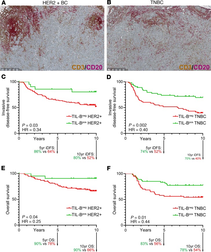
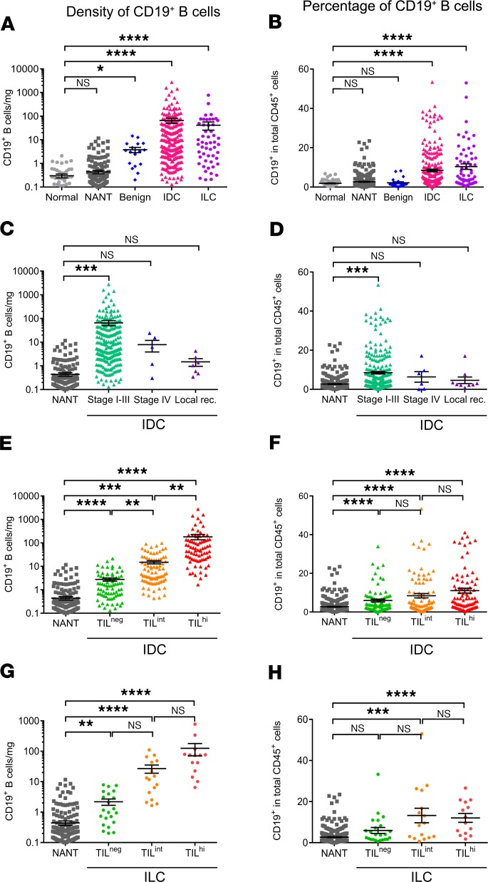
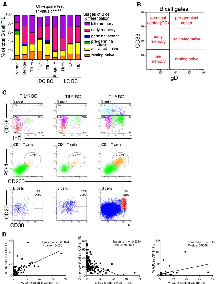
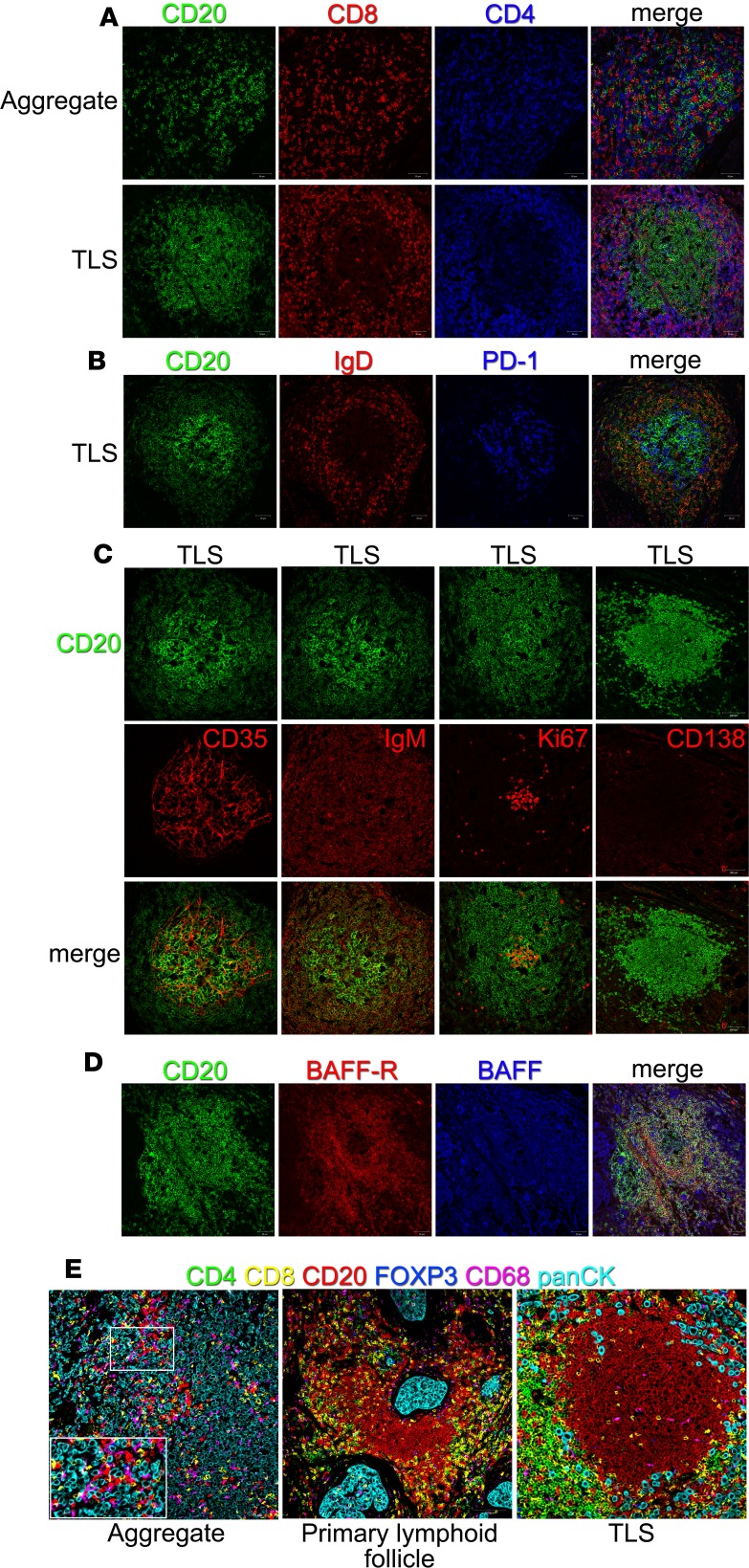
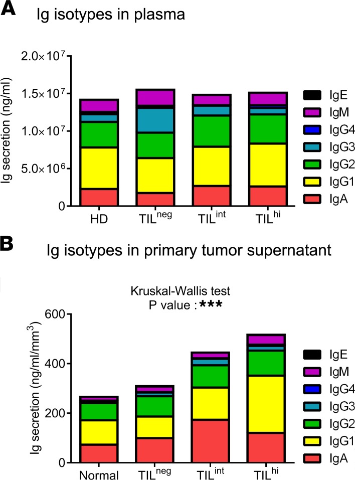
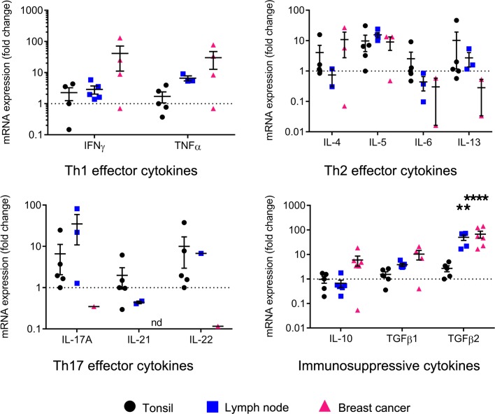
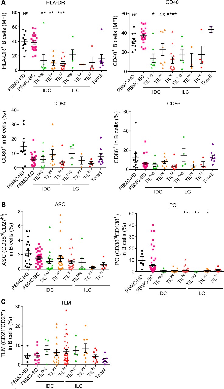
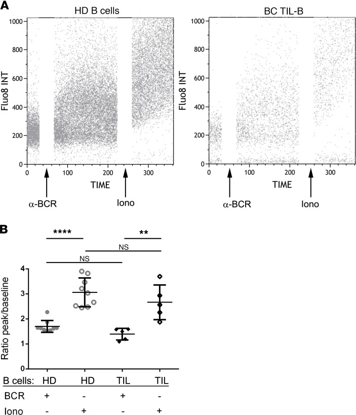
Tumor-infiltrating B-cells (TIL-B) in breast cancer (BC) have previously been associated with improved clinical outcomes; however, their role(s) in tumor immunity is not currently well known. This study confirms and extends the correlation between higher TIL-B densities and positive outcomes through an analysis of HER2-positive and triple-negative BC patients from the BIG 02-98 clinical trial (10yr mean follow-up). Fresh tissue analyses identify an increase in TIL-B density in untreated primary BC compared to normal breast tissues, which is associated with global, CD4+ and CD8+ TIL, higher tumor grades, higher proliferation and hormone receptor negativity. All B-cell differentiation stages are detectable but significant increases in memory TIL-B are consistently present. BC with higher infiltrates are specifically characterized by germinal center TIL-B, which in turn are correlated with TFH TIL and antibody-secreting TIL-B principally located in tertiary lymphoid structures. Some TIL-B also interact directly with tumor cells. Functional analyses reveal TIL-B are responsive to BCR stimulation ex vivo, express activation markers and produce cytokines and immunoglobulins despite reduced expression of the antigen-presenting molecules HLA-DR and CD40. Overall, these data support the concept that ongoing humoral immune responses are generated by TIL-B and help to generate effective anti-tumor immunity at the tumor site.

Keywords: Adaptive immunity; B cells; Breast cancer; Immunology; Oncology.


Conflict of interest statement

Conflict of interest: The authors have declared that no conflict of interest exists.

Figures

Figure 1
Figure 1. Prognostic value of tumor-infiltrating B cells in breast cancer.
(A and B) Representative sections of HER2 (A) and TN (B) breast cancer with extensive TIL stained with CD3/CD20. (CF) Kaplan-Meyer survival curves of 10-year invasive disease-free survival (iDFS) for 136 patients with HER2-positive disease (C), iDFS for 113 TN disease (D), overall survival (OS) for 136 patients with HER2-positive disease (E), and OS for 113 TN disease (F). Statistical analysis: log-rank (Mantel-Cox) test. See also Supplemental Tables 1 and 2.
Figure 2
Figure 2. B cell infiltration in breast cancer tissues.
(AH) B cell infiltration was determined as an absolute number normalized to the weight (mg) of the tissue sample (A, C, E, and G) and as a percentage (B, D, F, and H) of CD45+ TIL in the tissue by FACS. B cell infiltration was analyzed according to breast tissue type and cancer histology (A and B), BC stage (C and D), and global TIL infiltration in IDC (E and F) and in ILC (G and H). Data represent a combination of experiments involving individual patients and are displayed as the mean ± SEM by 1-way ANOVA with Dunn’s multiple comparisons test (normal, n = 62; NANT, n = 312; benign, n = 21; IDC, n = 241; ILC, n = 62; stage I–III, n = 241; stage IV, n = 6, local recurrence (rec.), n = 9; IDC TILneg, n = 78; IDC TILint, n = 84; IDC TILhi, n = 79; ILC TILneg, n = 21; ILC TILint, n = 16; ILC TILhi, n = 14). *P < 0.05; **P < 0.01; ***P < 0.001; ****P < 0.0001. NANT, nonadjacent nontumor; IDC, invasive ductal carcinoma; ILC, invasive lobular carcinoma; TIL, tumor-infiltrating lymphocytes. See also Supplemental Table 3 and Supplemental Figure 1.
Figure 3
Figure 3. Phenotypic characterization of TIL-B in breast cancer tissues.
(A and B) Stacked bars show the percentage of TIL-B subsets in total TIL-B according to the Bm classification (CD38 and IgD) (normal, n = 22; benign, n = 6; IDC TILneg, n = 9; IDC TILint, n = 27; IDC TILhi, n = 56; stage IV, n = 3; ILC TILneg, n = 4; ILC TILint, n = 10; ILC TILhi, n = 12). Data represent a combination of experiments involving individual patients and are displayed as the mean by χ2 test. ****P < 0.0001. (C) Representative dot plots show percentages of Bm subsets, TFH, and ASC in TILneg, TILint, and TILhi patients. (D) Graphs show the correlation between Bm3–4 and TFH (n = 57), Bm5 (n = 101), and ASC (n = 28). Data represent a combination of experiments involving individual patients and are displayed as single value by Spearman test. Bm, mature B cells; TFH, CD4+ T follicular helper; ASC, antibody-secreting B cells; recurrence; TIL, tumor-infiltrating lymphocytes; IDC, invasive ductal carcinoma; ILC, invasive lobular carcinoma. See also Supplemental Figure 2 and Supplemental Table 4.
Figure 4
Figure 4. B cell organization in breast cancer tissues.
(AD) Immunofluorescent staining of tumor-associated aggregates and TLS on FFPE sections (20× magnification). Tumor-infiltrating lymphocytes were revealed by CD20 (green), CD8 (red), and CD4 (blue) (A). Germinal centers were identified by the absence of naive IgD+ B cells (red) and the presence of PD-1+ cells (blue) (B), as well as CD35+ (red) follicular dendritic cells, IgM+ (red) memory B cells, Ki67+ (red) proliferating cells, and CD138+ (red) plasma cells (C). BAFF (blue) and its receptor (red) were detected in germinal center of breast cancer (D). (E) Representative multiplexed IHC images of breast cancer. Tumor-infiltrating lymphocytes were revealed by CD20 (red), CD8 (yellow), CD4 (green), FOXP3 (blue), and CD68 (magenta) and tumor cells by panCK (cyan) (20× magnification). See also Supplemental Figure 3.
Figure 5
Figure 5. Ig repertoire in breast cancer.
(A and B) Stacked bars quantify Ig subclasses in plasma from breast cancer patients and HD (A) and in breast tissue supernatant (B) (HD, n = 8; normal, n= 10; TILneg, n = 8; TILint, n = 4; TILhi, n = 9). Data represent a combination of experiments involving individual patients and are displayed as the mean ± SEM by 1-way ANOVA with Kruskal-Wallis test. ***P < 0.001. HD, healthy donors; TIL, tumor-infiltrating lymphocytes.
Figure 6
Figure 6. Cytokine profile of tumor-infiltrating B cells in breast cancer.
Scatter plots show the transcript levels of cytokines in sorted B cells from lymph node (n = 6) and BC (n = 6), normalized to MLN51, and relative to B cells from tonsil (n = 5). Data represent a combination of experiments involving individual patients and are displayed as mean ± SEM by 1-way ANOVA with Tukey multiple comparisons test. **P < 0.01; ****P < 0.0001. LN, lymph node; BC, breast cancer. See also Supplemental Figure 4.
Figure 7
Figure 7. Antigen-presenting cells functions of B cells in breast cancer.
(A) Scatter plots display the mean fluorescent intensity of HLA-DR and CD40, and the percentage of CD80 and CD86, on TIL-B (PBMC-HD, n = 10; PBMC-BC, n = 24; IDC TILneg, n = 8; IDC TILint, n = 8; IDC TILhi, n = 13; ILC TILneg, n = 5; ILC TILint, n = 3; ILC TILhi, n = 4; tonsil, n = 9). (B) Scatter plots display the percentage of ASC (PBMC-HD, n = 18; PBMC-BC, n = 33; IDC TILneg, n = 20; IDC TILint, n = 25; IDC TILhi, n = 21; ILC TILneg, n = 7; ILC TILint, n = 3; ILC TILhi, n = 5) and PC in B cells (PBMC-HD, n = 5; PBMC-BC, n = 63; IDC TILneg, n = 5; IDC TILint, n = 14; IDC TILhi, n = 36; ILC TILneg, n = 4; ILC TILint, n = 9; ILC TILhi, n = 8). (C) Scatter plot displays the percentage of tissue-like memory B cells using CD21CD27CD19+ in B cells (PBMC-HD, n = 5; PBMC-BC, n = 11; IDC TILneg, n = 5; IDC TILint, n = 16; IDC TILhi, n = 37; ILC TILneg, n = 5; ILC TILint, n = 8; ILC TILhi, n = 8; tonsil, n = 7). Data represent a combination of experiments involving individual patients and are displayed as mean ± SEM by 1-way ANOVA with Dunn’s multiple comparisons test. *P < 0.05; **P < 0.01; ***P < 0.001; ****P < 0.0001. PBMC, peripheral blood mononuclear cells; HD, healthy donors; BC, breast cancer; TIL, tumor-infiltrating lymphocytes; IDC, invasive ductal carcinoma; ILC, invasive lobular carcinoma; ASC, antibody-secreting cells; PC, plasma cells; TLM, tissue-like memory B cells.
Figure 8
Figure 8. BCR-mediated activation in tumor-infiltrating B cells.
(A and B) TIL-B were loaded with Fluo-8 and were BCR stimulated. Ionomycin was used as a positive control. (A) Representative dot plots show the calcium influx in B cells from HD and BC patients. (B) Scatter plot shows the ratio of calcium peak to baseline (HD, n = 9; TIL, n = 5). Data represent a combination of experiments involving individual patients and are displayed as mean ± SEM by 1-way ANOVA with Tukey multiple comparisons test. TIL, tumor-infiltrating lymphocytes; PBMC, peripheral blood mononuclear cells; BCR, B cell receptor; iono, ionomycin.

References

    1. David H. Rudolf Virchow and modern aspects of tumor pathology. Pathol Res Pract. 1988;183(3):356–364. doi: 10.1016/S0344-0338(88)80138-9. - DOI - PubMed
    1. Denkert C, et al. Tumor-associated lymphocytes as an independent predictor of response to neoadjuvant chemotherapy in breast cancer. J Clin Oncol. 2010;28(1):105–113. doi: 10.1200/JCO.2009.23.7370. - DOI - PubMed
    1. West NR, Milne K, Truong PT, Macpherson N, Nelson BH, Watson PH. Tumor-infiltrating lymphocytes predict response to anthracycline-based chemotherapy in estrogen receptor-negative breast cancer. Breast Cancer Res. 2011;13(6):R126. doi: 10.1186/bcr3072. - DOI - PMC - PubMed
    1. Loi S, et al. Prognostic and predictive value of tumor-infiltrating lymphocytes in a phase III randomized adjuvant breast cancer trial in node-positive breast cancer comparing the addition of docetaxel to doxorubicin with doxorubicin-based chemotherapy: BIG 02-98. J Clin Oncol. 2013;31(7):860–867. doi: 10.1200/JCO.2011.41.0902. - DOI - PubMed
    1. Adams S, et al. Prognostic value of tumor-infiltrating lymphocytes in triple-negative breast cancers from two phase III randomized adjuvant breast cancer trials: ECOG 2197 and ECOG 1199. J Clin Oncol. 2014;32(27):2959–2966. doi: 10.1200/JCO.2013.55.0491. - DOI - PMC - PubMed

Publication types