. 2019 Aug 13;129(11):4850-4862.
doi: 10.1172/JCI127471.

cGAS/STING axis mediates a topoisomerase II inhibitor-induced tumor immunogenicity

Affiliations

cGAS/STING axis mediates a topoisomerase II inhibitor-induced tumor immunogenicity

Zining Wang et al. J Clin Invest. .

Abstract

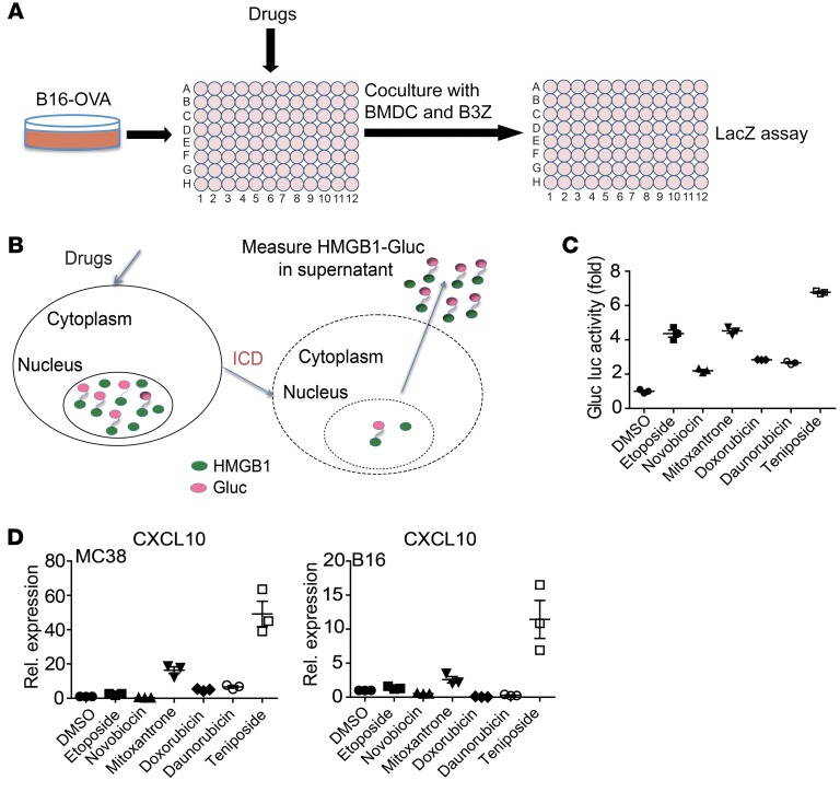
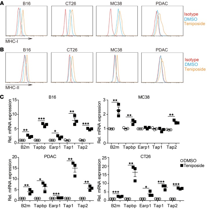
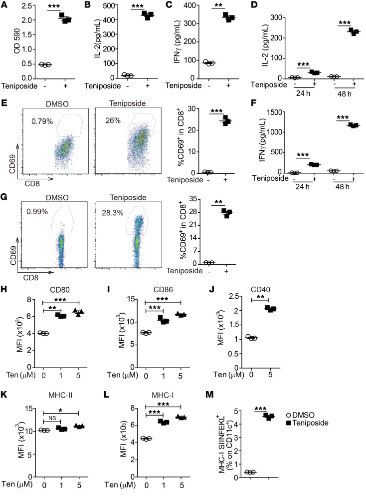
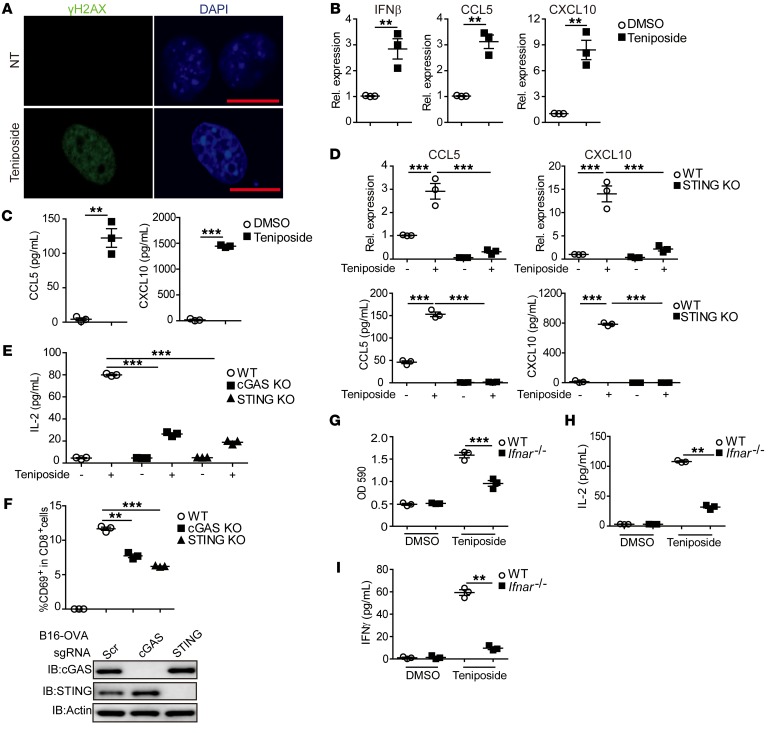
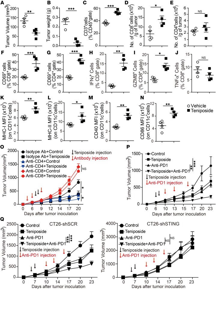
Checkpoint blockade antibodies have been approved as immunotherapy for multiple types of cancer, but the response rate and efficacy are still limited. There are few immunogenic cell death (ICD)-inducing drugs available that can kill cancer cells, enhance tumor immunogenicity, increase the in vivo immune infiltration, and thereby boosting a tumor response to immunotherapy. So far, the ICD markers have been identified as the few immuno-stimulating characteristics of dead cells, but whether the presence of such ICD markers on tumor cells translates into enhanced antitumor immunity in vivo is still investigational. To identify anticancer drugs that could induce tumor cell death and boost T cell response, we performed drug screenings based on both an ICD reporter assay and T cell activation assay. We identified that teniposide, a DNA topoisomerase II inhibitor, could induce high mobility group box 1 (HMGB1) release and type I interferon signaling in tumor cells, and teniposide-treated tumor cells could activate antitumor T cell response both in vitro and in vivo. Mechanistically, teniposide induced tumor cell DNA damage and innate immune signaling including NF-κB activation and STING-dependent type I interferon signaling, both of which contribute to the activation of dendritic cells and subsequent T cells. Furthermore, teniposide potentiated the antitumor efficacy of anti-PD1 on multiple types of mouse tumor models. Our findings showed that teniposide could trigger tumor immunogenicity, and enabled a potential chemo-immunotherapeutic approach to potentiate the therapeutic efficacy of anti-PD1 immunotherapy.

Keywords: Antigen presentation; Cancer immunotherapy; Immunology; Innate immunity; Oncology.


Conflict of interest statement

Conflict of interest: The authors have declared that no conflict of interest exists.

Figures

Figure 1
Figure 1. T cell–based drug screening identified ICD inducers.
(A) Outline of drug-screening protocol. B16-OVA tumor cells were seeded on 96-well plates and treated with drugs for 16 hours, then cocultured with BMDC and B3Z cells for 24 hours. LacZ reporter activity was measured as a surrogate marker for T cell activation. (B) Illustration of the principle of the HMGB1-Gluc reporter system. Once drugs or inhibitors induce tumor cell ICD, HMGB1-Gluc is released from the nucleus into the supernatant, and supernatant luciferase activity is detected. (C) MC38 (HMGB1-Gluc) cells were treated with different Top inhibitors or DMSO for 20 hours; then HMGB1-Gluc luciferase (Gluc luc) activity was measured. (D) MC38 and B16 cells were treated as in C, and then the mRNA expression level of CXCL10 was measured by qPCR. Rel., relative. Data in C and D are shown as mean ± SD of 3 independent experiments.
Figure 2
Figure 2. Teniposide induced ICD of tumor cells.
(A) MC38 (HMGB1-Gluc) and CT26 (HMGB1-Gluc) cells were treated with increasing doses of teniposide for 20 hours, and HMGB1-Gluc luciferase activity was measured. (B and C) CT26 cells were treated with teniposide or DMSO for 20 hours, and cell apoptosis (B) and surface expression of CRT(C) were detected by FACS. (D) CT26 tumor cells were pretreated with teniposide, etoposide, or freeze-thawed, followed by subcutaneous inoculation into BALB/c mice as a vaccine (n = 8 for control group with no tumor cell vaccine administered, teniposide group, and freeze-thawed group; n = 5 for etoposide group). After 8 days, mice were rechallenged with live CT26 cells. Shown are the percentages of tumor-free mice 30 days after rechallenge. Data in AC are shown as mean ± SD of 3 independent experiments. **P < 0.01; ***P < 0.001, 1-way ANOVA with Bonferroni’s post test (A), unpaired Student’s t test (B), log-rank (Mantel-Cox) test (D).
Figure 3
Figure 3. Teniposide enhanced expression of antigen-presenting machinery molecules on tumor cells.
(A and B) B16, MC38, PDAC, and CT26 cells were treated with teniposide or DMSO for 20 hours, and the surface expression of MHC-I and MHC-II was determined by FACS. (C) Cells were treated as in A, and the expression of antigen-presenting machinery genes were measured by qPCR. Data in A and B are shown as the representative results of 3 repeated experiments. Data in C are shown as mean ± SD of 3 independent experiments. *P < 0.05; **P < 0.01; ***P < 0.001, unpaired Student’s t test.
Figure 4
Figure 4. Teniposide-treated tumor cells induced T cell activation and DC maturation.
(AD) B16-OVA cells were treated with teniposide or DMSO for 16 hours, then cocultured with BMDC and B3Z cells for an additional 24 hours, after which B3Z activation was measured by LacZ activity, IL-2 production, and IFN-γ production (AC) and CD69 expression (D). (EG) B16-OVA cells were treated with teniposide or DMSO for 16 hours, then cocultures with BMDC and OT-I cells for an additional 24 hours or 48 hours, after which OT-I activation was measured by secretion of IL-2 and IFN-γ and surface expression of CD69. (HM) B16-OVA cells were treated with DMSO or indicated concentration of teniposide for 16 hours, then cocultured with BMDCs for an additional 24 hours, after which surface expression of CD80, CD86, CD40, MHC-II, MHC-I, and MHC-I–SIINFEKL on CD11c+ DCs was determined by FACS. Data in AM are shown as mean ± SD of 3 independent experiments. *P < 0.05; **P < 0.01; ***P < 0.001, unpaired Student’s t test (AG, J, M); 1-way ANOVA with Bonferroni’s post test (H, I, K, L).
Figure 5
Figure 5. Teniposide activated cGAS/STING-dependent IFN-I signaling in tumor cells.
(A) B16 cells were treated with teniposide or DMSO for 24 hours; then γH2AX expression was detected by immunofluorescence staining. Scale bar: 10 μm. (B) B16 cells were treated as in A; then the expression levels of IFN-β, CCL5, and CXCL10 were measured by qPCR. (C) Cells were treated as in A; then the supernatant levels of CCL5 and CXCL10 were measured by ELISA. (D) B16/WT and B16/STING KO cells were treated with teniposide or DMSO for 24 hours; then the levels of mRNA and protein expression of CCL5 and CXLC10 were measured by qPCR and ELISA, respectively. (E and F) B16-OVA/WT, B16-OVA/cGAS-KO and B16-OVA/STING-KO cells were treated with teniposide or DMSO for 16 hours, then cocultured with B3Z+BMDCs for an additional 24 hours. T cell activation was measured by supernatant IL-2 levels and surface expression of CD69. Protein expression of cGAS or STING was measured by Western blot. Actin was used as a loading control. (GI) B16-OVA cells were treated with teniposide or DMSO for 16 hours, then cocultured with B3Z in the presence of WT or Ifnar–/– BMDCs for an additional 24 hours, after which LacZ activity and the supernatant levels of IL-2 and IFN-γ were determined. Data shown in A are representative of 1 of 3 independent experiments. Data shown in BI are represented as mean ± SD of 3 independent experiment. **P < 0.01; ***P < 0.001, unpaired Student’s t test (B, C, GI); 1-way ANOVA with Bonferroni’s post test (DF).
Figure 6
Figure 6. Teniposide induced immune cell infiltration and potentiated efficacy of anti-PD1 therapy in a CT26 mouse tumor model.
(AN) Mice with established CT26 tumors were treated with teniposide or vehicle on days 6 and 7 (10 mg/kg, i.p.). Tumors were isolated on day 10, and tumor-infiltrating immune cells were analyzed by flow cytometry. Data are representative of 1 of 2 independent experiments. Shown are tumor volume (A), tumor weight (B), intratumoral T cells (C), numbers of tumor-infiltrating CD8+ T cells (D), CD4+ T cells (E), and expression of activation marker CD69 (F and G) and effector molecules IFN-γ, GZMB, and TNF-α (HJ) in CD8+ T cells. (KN) Surface expression levels of MHC-I, MHC-II, CD40, and CD86 on CD11c+ cells were determined by FACS. n = 4 mice per group. (O) Mice were injected with CD8 or CD4 depletion antibody on days 3, 6, and 9 after CT26 tumor inoculation, followed by teniposide treatment on days 7 and 8 (10 mg/kg, i.p.). Tumor volume is shown as mean ± SD. n = 5 per group. (P) Mice with established CT26 tumors were treated with teniposide, anti-PD1, or teniposide in combination with anti-PD1 at indicated time points. Tumor volume was shown as mean ± SD. n = 7 per group. (Q) Mice were inoculated with CT26-shSCR (scramble shRNA as control) or CT26-shSTING cells and then treated with indicated drugs. Tumor volume is shown as mean ± SD. n = 5 per group. *P < 0.05; **P < 0.01; ***P < 0.001, unpaired Student’s t test (AN) or 2-way ANOVA with Bonferroni’s post test (OQ).

References

    1. Topalian SL, et al. Immunotherapy: The path to win the war on cancer? Cell. 2015;161(2):185–186. doi: 10.1016/j.cell.2015.03.045. - DOI - PMC - PubMed
    1. Galluzzi L, Chan TA, Kroemer G, Wolchok JD, López-Soto A. The hallmarks of successful anticancer immunotherapy. Sci Transl Med. 2018;10(459):eaat7807. doi: 10.1126/scitranslmed.aat7807. - DOI - PubMed
    1. Zou W, Wolchok JD, Chen L. PD-L1 (B7-H1) and PD-1 pathway blockade for cancer therapy: Mechanisms, response biomarkers, and combinations. Sci Transl Med. 2016;8(328):328rv4. doi: 10.1126/scitranslmed.aad7118. - DOI - PMC - PubMed
    1. Yan L, Zhang W. Precision medicine becomes reality-tumor type-agnostic therapy. Cancer Commun (Lond) 2018;38(1):6. doi: 10.1186/s40880-018-0274-3. - DOI - PMC - PubMed
    1. Ansell SM, et al. PD-1 blockade with nivolumab in relapsed or refractory Hodgkin’s lymphoma. N Engl J Med. 2015;372(4):311–319. doi: 10.1056/NEJMoa1411087. - DOI - PMC - PubMed

Publication types

MeSH terms