. 2019 Nov;158(3):230-239.
doi: 10.1111/imm.13107. Epub 2019 Sep 11.

Immune evasion by Salmonella: exploiting the VPAC1/VIP axis in human monocytes

Affiliations

Immune evasion by Salmonella: exploiting the VPAC1/VIP axis in human monocytes

Basim Askar et al. Immunology. 2019 Nov.

Abstract

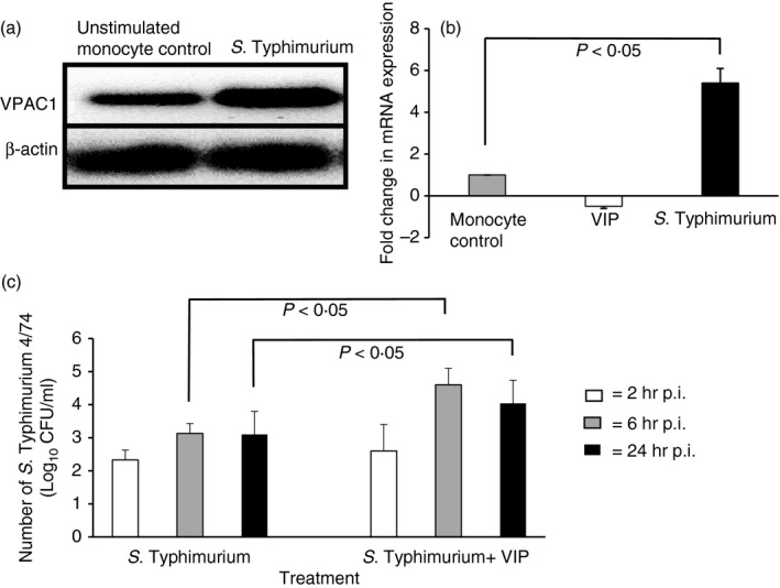
Immune evasion is a critical survival mechanism for bacterial colonization of deeper tissues and may lead to life-threatening conditions such as endotoxaemia and sepsis. Understanding these immune evasion pathways would be an important step for the development of novel anti-microbial therapeutics. Here, we report a hitherto unknown mechanism by which Salmonella exploits an anti-inflammatory pathway in human immune cells to obtain survival advantage. We show that Salmonella enterica serovar Typhimurium strain 4/74 significantly (P < 0·05) increased expression of mRNA and surface protein of the type 1 receptor (VPAC1) for anti-inflammatory vasoactive intestinal peptide (VIP) in human monocytes. However, we also show that S. Typhimurium induced retrograde recycling of VPAC1 from early endosomes to Rab11a-containing sorting endosomes, associated with the Golgi apparatus, and anterograde trafficking via Rab3a and calmodulin 1. Expression of Rab3a and calmodulin 1 were significantly increased by S. Typhimurium infection and W-7 (calmodulin antagonist) decreased VPAC1 expression on the cell membrane while CALP-1 (calmodulin agonist) increased VPAC1 expression (P < 0·05). When infected monocytes were co-cultured with VIP, a significantly higher number of S. Typhimurium were recovered from these monocytes, compared with S. Typhimurium recovered from monocytes cultured only in cell media. We conclude that S. Typhimurium infection exploits host VPAC1/VIP to gain survival advantage in human monocytes.

Keywords: Salmonella; Rab; calmodulin; immune evasion; vasoactive intestinal peptide.


Figures

Figure 1
Figure 1
Increased survival of Salmonella enterica serovar Typhimurium 4/74 in human monocytes co‐cultured with vasoactive intestinal peptide (VIP) is associated with increased expression of VIP receptor 1 (VPAC1). (a) Reverse transcription PCR showing constitutive and S. Typhimurium‐induced expression of VPAC1 in monocytes after 6 hr post‐infection (p.i.). Comparison between VPAC1 expression is also shown with the reference gene β‐actin, which is unchanged in both uninfected and infected monocytes. (b) Quantitative PCR showing expression of VPAC1 after 6 hr p.i. in monocytes infected with S. Typhimurium or cultured with VIP (10−7 m). Fold changes are expressed in comparison with mRNA expression measured in unstimulated (control) monocytes, assigned an arbitrary expression value of 1. (c) Recovery of S. Typhimurium from monocytes cultured with or without VIP (10−7 m) at different times p.i.: white bar = 2 hr p.i., grey bar = 6 hr p.i., black bar = 24 hr p.i. Histograms (b and c) show means calculated from five separate experiments performed in triplicate. Connecting bars show significant differences at P = 0·05, error bars show standard deviation from the mean.
Figure 2
Figure 2
Salmonella enterica serovar Typhimurium 4/74 increases vasoactive intestinal peptide receptor 1 (VPAC1) protein in the cytosol and cell membrane of human monocytes. VPAC1 expression is shown in representative FACS analyses of five independent experiments performed in (a–e) the cytoplasm (permeabilized) and (f–j) cell membrane (non‐permeabilized) at 6 hr post‐infection (p.i.). In all cases, lower left quadrants were set according to fluorescence expressed by monocytes incubated with secondary antibody only (control). Histograms show mean population sizes expressing VPAC1 after each treatment; permeabilized monocytes (e) and non‐permeabilized monocytes (j) calculated from five independent experiments performed in triplicate. Connecting bars show significant differences at P = 0·01 to P = 0·05, error bars show standard deviation from the mean.
Figure 3
Figure 3
Salmonella enterica serovar Typhimurium infection stimulates retrograde transport of vasoactive intestinal peptide receptor 1 (VPAC1) from early/sorting endosome to Rab11a‐containing recycling endosome localized with the trans‐Golgi network in human monocytes. Confocal laser scanning microscopy showing co‐localization of VPAC1 with endosome antigen 1 (EEA1) and Rab11a in human monocytes, each panel shows a merged plane image 4 μm below the cell surface. (a) Co‐localization of VPAC1 immunofluorescence (green Alexa 488) with early endosome antigen 1 (red Alexa 647) (white arrows) with increased levels of VPAC1/EEA1 association in S. Typhimurium‐infected monocytes at 6 hr postinfection. (b) Co‐localization of VPAC1 (green Alexa 488) with Rab11a (red Alexa 647) (white arrows) is shown only in S. Typhimurium‐infected cells, and in (c) co‐localization of Rab11a (red Alexa 647) with TGN46 (green Alexa 488) (white arrows) is shown only in S. Typhimurium‐infected cells. Confocal laser scanning microscopy images shown are representative of five independent experiments performed in triplicate. Scale bar (bottom left) = 20 μm.
Figure 4
Figure 4
Salmonella enterica serovar Typhimurium 4/74 induces packaging of vasoactive intestinal peptide receptor 1 (VPAC1) within Rab3a/CAM1 containing secretory vesicles for anterograde transport to the cell membrane of human monocytes. (a, b) Confocal laser scanning microscopy showing co‐localization of VPAC1 with Rab3a and CAM1. Each panel shows a merged plane image 4 μm below the cell surface. (a) Co‐localization of VPAC1 (green Alexa 488) with Rab3a (red Alexa 647) (white arrows) in S. Typhimurium‐infected monocytes. (b) Co‐localization of CAM1 (green Alexa 488) with Rab3a (red Alexa 647) (white arrows) in S. Typhimurium‐infected monocytes. Images shown are representative of five independent experiments performed in triplicate. Scale bar (bottom left) = 20 μm. (c) Quantitative PCR showing increased expression of Rab3a and CAM1 mRNA in monocytes 6 hr postinfection with S. Typhimurium. (d) Quantitative PCR showing fold changes in VPAC1 mRNA with or without CAM1 agonist (CALP1) or antagonist (W‐7). Each bar is a mean of five independent experiments performed in triplicate. White bar = without agonist/antagonist, black bar = + CALP1 (agonist), grey bar = + W‐1 (antagonist). Connecting bars show significant differences at P = 0·05, error bars show standard deviation from the mean. Fold changes are expressed as a comparison with mRNA expression measured in unstimulated (control) monocytes after 6 hr, assigned an arbitrary expression value of 1.
Figure 5
Figure 5
CAM1 increases vasoactive intestinal peptide receptor 1 (VPAC1) protein expression on the cell membrane of monocytes infected with Salmonella enterica serovar Typhimurium 4/74. (a–j) FACS analysis showing VPAC1 expression on the monocyte membrane 6 hr postinfection with S. Typhimurium or monocytes cultured with vasoactive intestinal peptide (VIP; 10−7 m) for 6 hr, with or without CAM1 agonist (CALP1; 20 μg/ml) or CAM1 antagonist (W‐7; 30 μg/ml). FACS analyses shown are representative of experiments performed in triplicate on five separate occasions. y‐axes, SSC = Side scatter; x‐axis, VPAC1 expression. Quantitative analysis of experimental means are shown in (k). Black bars = 2 hr post‐treatment/infection, white bars = 6 hr post‐treatment/infection and grey bars = 24 hr post‐treatment/infection. Connecting bars show significant differences at P = 0·05, error bars show standard deviation from the mean. (l) Diagrammatic representation of S. Typhimurium‐induced trafficking of VPAC1 receptors.(1) S. Typhimurium invades monocyte. (2) VPAC1 is packaged in the EEA1‐positive sorting endosome (SE). (3) Retrograde transport of VPAC1 occurs via Rab11a‐positive recycling endosome (RE), which is associated with TGN46 on the Golgi apparatus. (4) Anterograde transport of VPAC1 occurs via Rab3a/CAM1 positive secretory vesicle (SV).

References

    1. Annane D, Bellissant E, Cavaillon JM. Septic shock. Lancet 2005; 365:63–78. - PubMed
    1. Davis BH. Improved diagnostic approaches to infection/sepsis detection. Expert Rev Mol Diagn 2005; 2005:193–207. - PubMed
    1. Ward NS, Casserly B, Ayala A. The compensatory anti‐inflammatory response syndrome (CARS) in critically ill patients. Clin Chest Med 2008; 29:617–25. - PMC - PubMed
    1. Danikas DD, Karakantza M, Theodorou GL, Sakellaropoulos GC, Gogos CA. Prognostic value of phagocytic activity of neutrophils and monocytes in sepsis. Correlation to CD64 and CD14 antigen expression. Clin Exp Immunol 2008; 154:87–97. - PMC - PubMed
    1. Cohen J. Sepsis and septic shock: inching forwards. Clin Med 2009; 9:256–7. - PMC - PubMed

Publication types

MeSH terms

Substances

LinkOut - more resources