

doi: 10.1016/j.jchemneu.2019.101661.
Epub 2019 Aug 10.
Monosynaptic tracing: a step-by-step protocol
Affiliations
- PMID: 31408693
- PMCID: PMC11526806
- DOI: 10.1016/j.jchemneu.2019.101661
Item in Clipboard
Monosynaptic tracing: a step-by-step protocol
J Chem Neuroanat.
2019 Dec.
Abstract
Monosynaptic tracing using deletion-mutant rabies virus allows whole-brain mapping of neurons that are directly presynaptic to a targeted population of neurons. The most common and robust way of implementing it is to use Cre mouse lines in combination with Cre-dependent adeno-associated viral vectors for expression of the required genes in the targeted neurons before subsequent injection of the rabies virus. Here we present a step-by-step protocol for performing such experiments using first-generation (ΔG) rabies viral vectors.
Copyright © 2019. Published by Elsevier B.V.
Figures
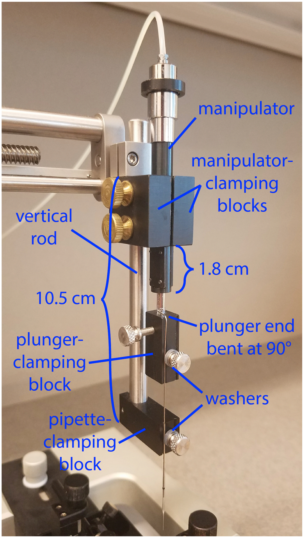
The injection apparatus consists of a manual hydraulic manipulator that advances a plunger through the bore of a pulled glass pipette. Three of the four custom parts (see Supplementary File 1 for fabrication information) are mounted on the vertical rod that comes with the stereotax and hold both the micromanipulator and the pipette; the fourth is mounted on the moveable shaft of the manipulator and holds the plunger that moves within the bore of the pipette to expel the virus. For best results, adhere to the positioning of the components shown here. The vertical metal rod extends approximately 10.5 cm below the metal clamp that holds the vertical rod to the stereotax arm, and the bottom of the pipette-clamping block is attached to the vertical rod (with a 4–40 screw) and flush with the end of it. The two symmetrical micromanipulator-clamping blocks are clamped (with two brass 10–32 screws) to both the vertical rod and the micromanipulator; the blocks should be flush against the metal clamp, and the end of the micromanipulator shaft is ~1.8 cm below the bottom edge of the clamping blocks. The plunger-clamping block is attached (with a 4–40 screw) to the piston of the micromanipulator, with the grooves of the pipette-clamping block and those of the plunger-clamping block aligned so that the plunger is in line with the pipette. The two 4–40 thumbscrews that pin the pipette and its plunger into the grooves in their respective blocks have nylon washers to provide grip and prevent breakage of the glass. While the exact positioning of the custom parts on the stereotax rod is not critical, the positions shown here work well, and major deviation from them could result in the inability to raise the pipette high enough to allow the stereotax arm to be swung out of the way when inserting and removing the mouse or, conversely, the inability to lower the pipette to the desired depth in the brain.
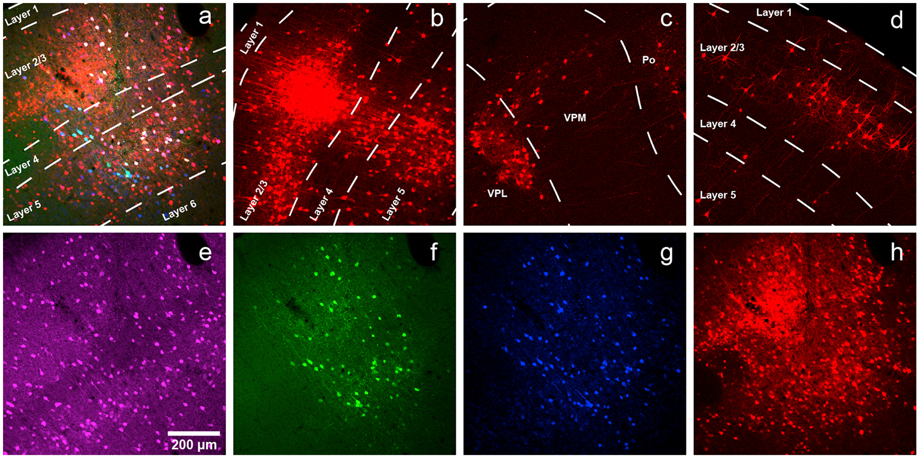
a) Injection site in primary somatosensory cortex (coordinates: AP= −0.58 mm with respect to bregma, LM = 3.00 mm with respect to bregma, DV = −1.00 mm with respect to the surface of the brain) of a PV-Cre mouse injected with a mixture of AAV1-syn-FLEX-splitTVA-EGFP-tTA and AAV1-TREtight-mTagBFP2-B19 G, followed by RVΔG-4mCherry(EnvA) a week later. Green = anti-EGFP staining, blue = mTagBFP2, red = mCherry. Individual channels from this field are shown in panels e–h. Scale bar: 200 μm, applies to all panels. b) Labeled presynaptic neurons in ipsilateral secondary somatosensory cortex. The dense central region is not an injection site but instead just a concentrated group of transsynaptically-labeled neurons. c) Labeled presynaptic neurons in ipsilateral thalamus (VPL, VPM and Po). d) Labeled presynaptic neurons in contralateral S1. e–h) Individual channels from the field shown in panel a. e) anti-parvalbumin staining (not shown in panel a). f) anti-EGFP staining, indicating expression from the first, Cre-dependent AAV (note that the EGFP signal is not usually visible when the helper viruses are used at the recommended dilutions). g) mTagBFP2, indicating expression from the second, tTA-dependent AAV. h) mCherry, marking activity of the rabies virus.
References
-
- Ahrlund-Richter S, Xuan Y, van Lunteren JA, Kim H, Ortiz C, Pollak Dorocic I, Meletis K, Carlen M, 2019. A whole-brain atlas of monosynaptic input targeting four different cell types in the medial prefrontal cortex of the mouse. Nat. Neurosci 22, 657–668. - PubMed
Publication types
MeSH terms
Substances
Grants and funding
LinkOut - more resources
Full Text Sources
Research Materials
