

Leading progress in heart regeneration and repair
- PMID: 31408771
- PMCID: PMC7376987
- DOI: 10.1016/j.ceb.2019.07.005
Leading progress in heart regeneration and repair
Abstract
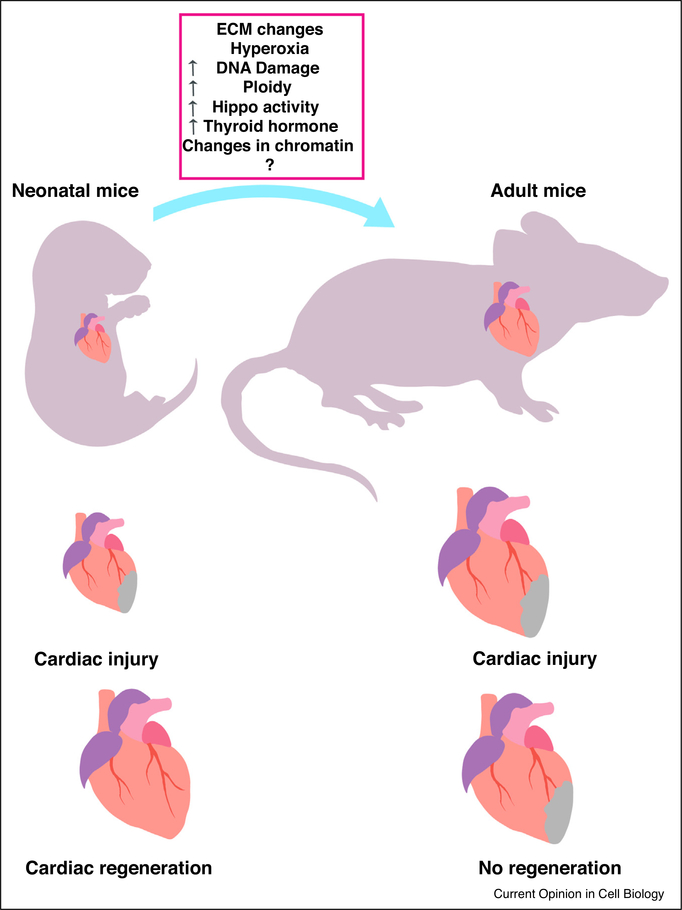
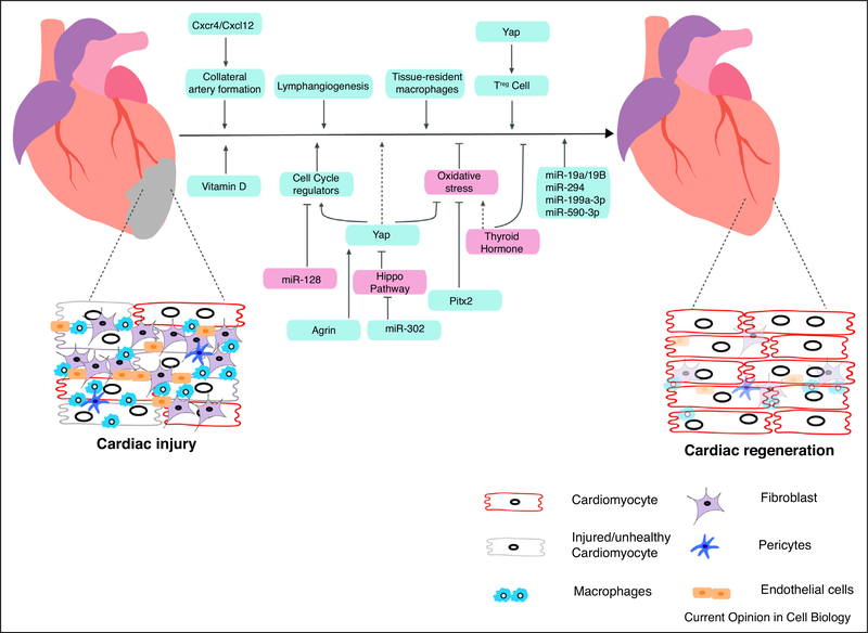
Ischemic heart disease is one of the leading causes of mortality. Myocardial infarction causes loss of cardiomyocytes in the injury area accompanied by formation of a fibrotic scar. This initiates a cascade of events including further loss of myocyte, increased fibrosis, and pathological cardiac hypertrophy, eventually leading to the heart failure. Cardiomyocytes in mammals have limited regenerative potential due to post mitotic nature of cardiomyocytes. Recently, multiple studies have provided substantial insights in to the molecular pathways governing this block in adult cardiomyocyte proliferation, and successfully employed that understanding to achieve cardiac regeneration. These strategies include directly reprograming the cardiomyocytes or manipulating the cardiac interstitium to repair the injured heart. In this review, we discuss the recent advances made in the field in the past two years.
Copyright © 2019 Elsevier Ltd. All rights reserved.
Conflict of interest statement
Conflict of interest statement
None declared.
Figures
References
-
- Leach JP, Martin JF: Cardiomyocyte proliferation for therapeutic regeneration. Curr Cardiol Rep 2018, 20:63. - PubMed
-
- Bergmann O, Zdunek S, Felker A, Salehpour M, Alkass K, Bernard S, Sjostrom SL, Szewczykowska M, Jackowska T, Remedios C et al.: Dynamics of cell generation and turnover in the human heart. Cell 2015, 161:1566–1575. - PubMed
-
- Ye L, D’Agostino G, Loo S, Wang C, Su L, Tan S, Tee G, Pua C, Pena E, Belen R et al.: Early regenerative capacity in the porcine heart. Circulation 2018, 138 CIRCULATIONAHA.117.031542. - PubMed
Publication types
MeSH terms
Grants and funding
LinkOut - more resources
Full Text Sources
Miscellaneous
