Review
. 2019 Aug 12;20(16):3913.
doi: 10.3390/ijms20163913.

Roles of Myosin-Mediated Membrane Trafficking in TGF-β Signaling

Affiliations
Review

Roles of Myosin-Mediated Membrane Trafficking in TGF-β Signaling

Chih-Ling Chung et al. Int J Mol Sci. .

Abstract

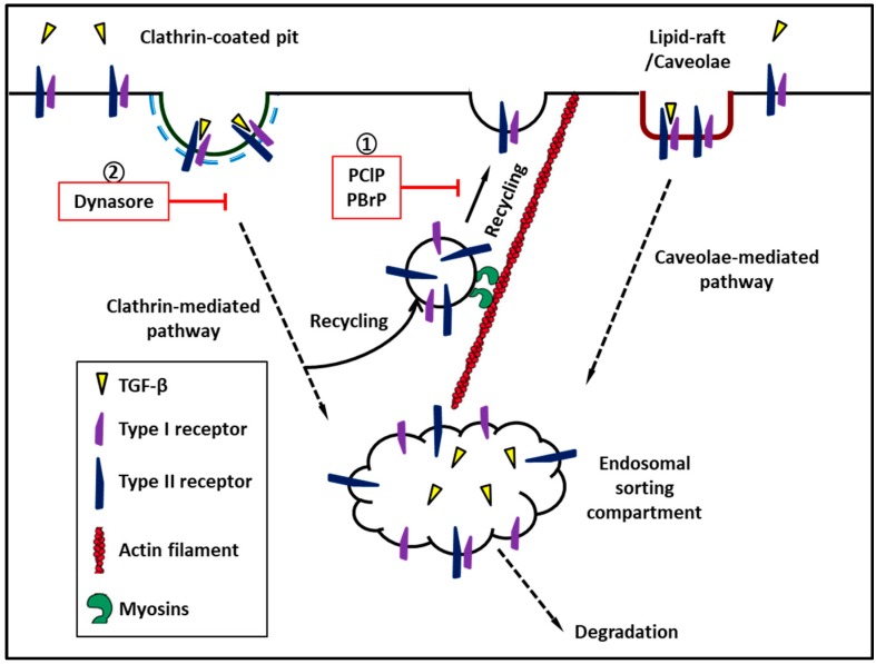
Recent findings have revealed the role of membrane traffic in the signaling of transforming growth factor-β (TGF-β). These findings originate from the pivotal function of TGF-β in development, cell proliferation, tumor metastasis, and many other processes essential in malignancy. Actin and unconventional myosin have crucial roles in subcellular trafficking of receptors; research has also revealed a growing number of unconventional myosins that have crucial roles in TGF-β signaling. Unconventional myosins modulate the spatial organization of endocytic trafficking and tether membranes or transport them along the actin cytoskeletons. Current models do not fully explain how membrane traffic forms a bridge between TGF-β and the downstream effectors that produce its functional responsiveness, such as cell migration. In this review, we present a brief overview of the current knowledge of the TGF-β signaling pathway and the molecular components that comprise the core pathway as follows: ligands, receptors, and Smad mediators. Second, we highlight key role(s) of myosin motor-mediated protein trafficking and membrane domain segregation in the modulation of the TGF-β signaling pathway. Finally, we review future challenges and provide future prospects in this field.

Keywords: TGF-β; clathrin-coated pits; endocytosis; lipid-rafts; subcellular trafficking; unconventional myosin.


Conflict of interest statement

The authors declare no conflict of interest.

Figures

Figure 1
Figure 1
Bar diagrams showing the overall architectures of human myosins discussed in this review. All the myosins have conserved motor heads (blue), different numbers of IQ motifs (gray) followed by a distinct tails with various functional domains. Colored boxes represent different regions predicted by sequence homology: PH, pleckstrin homology domain; TH1, TH2, and TH3, tail homology region 1, 2, and 3; CC, coiled-coil domains; GTD, globular tail domain; CBD, cargo binding domain; SH3, Src-homology 3 domain. Myosin-Va, and myosin-X exist as constitutive dimers and myosin-VI may exist in both monomeric and dimeric forms.
Figure 2
Figure 2
Myosins regulate recycling of transforming growth factor-β receptor to cell surface. ① Pentachloropseudilin and pentabromopseudilin suppress recycle of TGF-β receptor to plasma membrane. ② Dynasore, a cell-permeable inhibitor of dynamin which inhibits clathrin-mediated endocytosis of TGF-β receptors.

References

    1. Kingsley D.M. The tgf-beta superfamily: New members, new receptors, and new genetic tests of function in different organisms. Genes Dev. 1994;8:133–146. doi: 10.1101/gad.8.2.133. - DOI - PubMed
    1. Rifkin D.B. Latent transforming growth factor-beta (tgf-beta) binding proteins: Orchestrators of tgf-beta availability. J. Biol. Chem. 2005;280:7409–7412. doi: 10.1074/jbc.R400029200. - DOI - PubMed
    1. Massagué J., Wotton D. Transcriptional control by the tgf-β/smad signaling system. Embo. J. 2000;19:1745–1754. doi: 10.1093/emboj/19.8.1745. - DOI - PMC - PubMed
    1. Shi Y., Massagué J. Mechanisms of tgf-β signaling from cell membrane to the nucleus. cell. 2003;113:685–700. doi: 10.1016/S0092-8674(03)00432-X. - DOI - PubMed
    1. Derynck R., Zhang Y.E. Smad-dependent and smad-independent pathways in tgf-β family signalling. Nature. 2003;425:577. doi: 10.1038/nature02006. - DOI - PubMed

LinkOut - more resources