. 2019 Aug 12;8(8):877.
doi: 10.3390/cells8080877.

Inhibition of the Adenosine A2A Receptor Mitigates Excitotoxic Injury in Organotypic Tissue Cultures of the Rat Cochlea

Affiliations

Inhibition of the Adenosine A2A Receptor Mitigates Excitotoxic Injury in Organotypic Tissue Cultures of the Rat Cochlea

Belinda Rx Han et al. Cells. .

Abstract

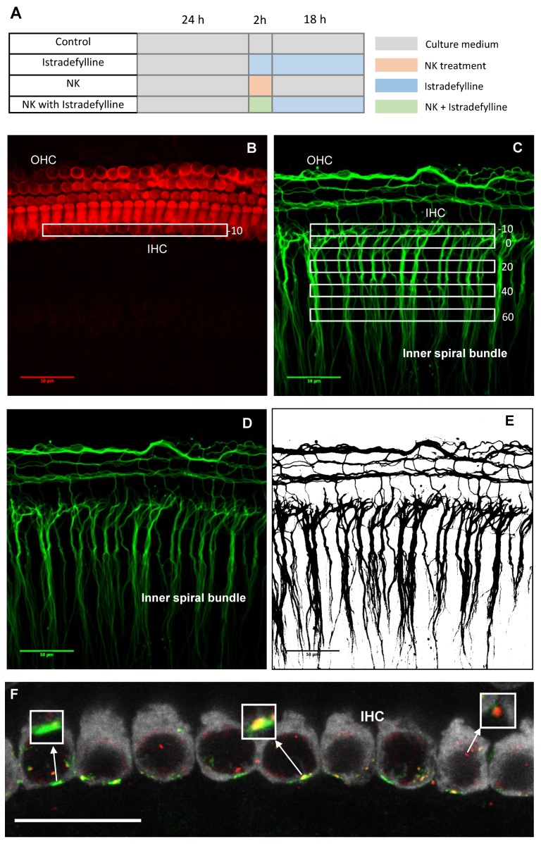
The primary loss of cochlear glutamatergic afferent nerve synapses due to noise or ageing (cochlear neuropathy) often presents as difficulties in speech discrimination in noisy conditions (hidden hearing loss (HHL)). Currently, there is no treatment for this condition. Our previous studies in mice with genetic deletion of the adenosine A2A receptor (A2AR) have demonstrated better preservation of cochlear afferent synapses and spiral ganglion neurons after noise exposure compared to wildtype mice. This has informed our current targeted approach to cochlear neuroprotection based on pharmacological inhibition of the A2AR. Here, we have used organotypic tissue culture of the Wistar rat cochlea at postnatal day 6 (P6) to model excitotoxic injury induced by N-methyl-d-aspartate (NMDA)/kainic acid (NK) treatment for 2 h. The excitotoxic injury was characterised by a reduction in the density of neural processes immediately after NK treatment and loss of afferent synapses in the presence of intact sensory hair cells. The administration of istradefylline (a clinically approved A2AR antagonist) reduced deafferentation of inner hair cells and improved the survival of afferent synapses after excitotoxic injury. This study thus provides evidence that A2AR inhibition promotes cochlear recovery from excitotoxic injury, and may have implications for the treatment of cochlear neuropathy and prevention of HHL.

Keywords: adenosine A2A receptor; cochlear explant; cochlear synaptopathy; glutamate excitotoxicity; hidden hearing loss; istradefylline.


Conflict of interest statement

The authors declare no conflict of interest.

Figures

Figure 1
Figure 1
(A) Outline of the study. (B) The regions of interest (−10 to +60 μm) at the inner hair cell level (myosin VIIa immunolabelling) and (C) at the inner spiral bundle level (β-tubulin immunolabelling) in organotypic tissue cultures of the P6 Wistar rat cochlea. (D) An original projected image of neurites immunolabelled with β-tubulin and (E) the converted binary image used for data analysis. (F) An image of the inner hair cell synaptic region: a paired synapse (yellow), orphan postsynaptic density protein (PSD95, green) and orphan presynaptic ribbon (CtBP2, red). Scale bars (BE), 50 μm; (F) 20 μm.
Figure 2
Figure 2
The effect of excitoxic injury in the neonatal rat cochlea induced by co-administration of N-methyl-d-aspartate (NMDA) and kainic acid (NK treatment). Images B–E show the NK effect in the function of time in order to find the best post-NK time point for further experiments in the study. (A) P6 Wistar rat cochlea labelled with Myosin VIIa (red; sensory hair cells) and β-tubulin (green; neurites of the spiral ganglion neurons) in normal culture medium; (B) Immediately after incubation with NK; (C) 6 h; (D) 18 h; (E) 24 h after NK treatment. Images are representative of five experiments per time point. Scale bars, 50 μm.
Figure 3
Figure 3
The effect of istradefylline treatment on cochlear explants exposed to excitotoxic injury. (A) P6 Wistar rat cochleae labelled with Myosin VIIa (red; sensory hair cells) and β-tubulin (green; spiral ganglion neurons and their neurites) in normal culture medium at low magnification (20×). The white box indicates the region of interest. (B) Cochlea cultured in normal medium (n = 7) and (C) culture medium containing istradefylline (n = 5). (D) Rat cochlea 18 h after NK-induced injury (n = 7) and (E) with istradefylline treatment (NK + Istradefylline; n = 10). IHC, inner hair cells; OHC, outer hair cells; SGN, spiral ganglion neurons. Images are representative of the number of experiments shown in brackets for each group. Scale bars, (A) 100 μm; (BE) 50 μm.
Figure 4
Figure 4
The relative density of SGN neurites in cochlear explants at different regions of interest designated as (A) −10 μm, (B) 0 μm, (C) +20 μm, (D) +40 μm and (E) +60 μm. CM, normal culture medium (n = 7); Istra, istradefylline (n = 5); NMDA/kainic acid, NK (n = 7), NKIstra, NK + istradefylline (n = 10). Data presented as mean ± SEM. * p < 0.05; ** p < 0.01; *** p < 0.001, one-way ANOVA followed by a post-hoc Sidak’s multiple comparisons test.
Figure 5
Figure 5
Ribbon synapses in the mid-cochlear region of P6 Wistar rats. Cochlear tissues immunolabelled with PSD95 (postsynaptic density protein, green), CtBP2 (synaptic ribbons, red) and myosin VIIa (inner hair cells, grey) antibodies in (A) normal culture medium (CM); (B) with addition of istradefylline (Istra); (C) with NMDA/kainic acid (NK); (D) a combined NKIstra treatment. Scale bars, 20 μm. (E) Number of paired synapses per inner hair cell in different treatment groups. Data presented as mean ± SEM. CM, n = 5; Istra, n = 5; NK, n = 4, NKIstra, n = 6. ** p < 0.01; *** p < 0.001, one-way ANOVA followed by a post-hoc Sidak’s multiple comparisons test. Images A–D are representative of the number of experiments shown for each group.
Figure 5
Figure 5
Ribbon synapses in the mid-cochlear region of P6 Wistar rats. Cochlear tissues immunolabelled with PSD95 (postsynaptic density protein, green), CtBP2 (synaptic ribbons, red) and myosin VIIa (inner hair cells, grey) antibodies in (A) normal culture medium (CM); (B) with addition of istradefylline (Istra); (C) with NMDA/kainic acid (NK); (D) a combined NKIstra treatment. Scale bars, 20 μm. (E) Number of paired synapses per inner hair cell in different treatment groups. Data presented as mean ± SEM. CM, n = 5; Istra, n = 5; NK, n = 4, NKIstra, n = 6. ** p < 0.01; *** p < 0.001, one-way ANOVA followed by a post-hoc Sidak’s multiple comparisons test. Images A–D are representative of the number of experiments shown for each group.
Figure 6
Figure 6
Confocal images of the P6 Wistar rat cochlea showing adenosine A2A receptor (A2AR) distribution at the synaptic region (SR) of the inner hair cells (arrows). (A) A2aR immunofluorescence (red), Myosin VIIa immunolabelling of the sensory hair cells (green) and β-tubulin labelling of SGN neurites (blue). (B) Peptide block control. Images are representative of eight cochleae obtained from four animals. Scale bars, 20 μm.

References

    1. Bohne B.A., Harding G.W. Degeneration in the cochlea after noise damage: primary versus secondary events. Am. J. Otol. 2000;21:505–509. - PubMed
    1. Kujawa S.G., Liberman M.C. Adding insult to injury: cochlear nerve degeneration after “temporary” noise-induced hearing loss. J. Neurosci. 2009;29:14077–14085. doi: 10.1523/JNEUROSCI.2845-09.2009. - DOI - PMC - PubMed
    1. Kujawa S.G., Liberman M.C. Synaptopathy in the noise-exposed and ageing cochlea: Primary neural degeneration in acquired sensorineural hearing loss. (Pt B)Hear. Res. 2015;330:191–199. doi: 10.1016/j.heares.2015.02.009. - DOI - PMC - PubMed
    1. Fernandez K.A., Jeffers P.W., Lall K., Liberman M.C., Kujawa S.G. Ageing after noise exposure: acceleration of cochlear synaptopathy in “recovered” ears. J. Neurosci. 2015;35:7509–7520. doi: 10.1523/JNEUROSCI.5138-14.2015. - DOI - PMC - PubMed
    1. Viana L.M., O’Malley J.T., Burgess B.J., Jones D.D., Oliveira C.A., Santos F., Merchant S.N., Liberman L.D., Liberman M.C. Cochlear neuropathy in human presbycusis: Confocal analysis of hidden hearing loss in post-mortem tissue. Hear. Res. 2015;327:78–88. doi: 10.1016/j.heares.2015.04.014. - DOI - PMC - PubMed

Publication types