. 2019 Aug 12;9(8):358.
doi: 10.3390/biom9080358.

Anti-Angiogenic Effect of Asperchalasine A Via Attenuation of VEGF Signaling

Affiliations

Anti-Angiogenic Effect of Asperchalasine A Via Attenuation of VEGF Signaling

Jun Yeon Park et al. Biomolecules. .

Abstract

Cytochalasans are a group of structurally diverse fungal polyketide-amino acid hybrid metabolites that exhibit diverse biological functions. Asperchalasine A was identified and isolated from an extract of the marine-derived fungus, Aspergillus. Asperchalasine A is a cytochalasan dimer which consists of two cytochalasan molecules connected by an epicoccine. This study investigated the potential antiangiogenic effects of Aspergillus extract and asperchalasine A, which significantly inhibited cell adhesion and tube formation in human umbilical vein endothelial cells (HUVECs). Aspergillus extract and asperchalasine A decreased the vascular endothelial growth factor (VEGF) and vascular endothelial growth factor receptor (VEGFR)-2 mRNA expression in a concentration-dependent manner. In addition, Aspergillus extract and asperchalasine A inhibited angiogenesis via downregulation of VEGF, p-p38, p-extracellular signal-regulated protein kinase (ERK), p-VEGFR-2, and p-Akt signaling pathways. Moreover, Aspergillus extract and asperchalasine A significantly inhibited the amount of blood vessel formation in fertilized chicken eggs using a chorioallantoic membrane assay. Our results provide experimental evidence of this novel biological activity of the potential antiangiogenic substances, Aspergillus extract, and asperchalasine A. This study also suggests that Aspergillus extract and its active component asperchalasine A are excellent candidates as adjuvant therapeutic substances for cancer prevention and treatment.

Keywords: HUVEC; VEGF; angiogenesis; asperchalasine A; metastasis.


Conflict of interest statement

The authors declare no conflict of interest.

Figures

Figure 1
Figure 1
The effect of Aspergillus extract and asperchalasine A on human umbilical vein endothelial cells’ (HUVECs) viability. (A) Chemical structure of asperchalasine A. (B,C) Cells were treated with Aspergillus extract and asperchalasine A in a series of concentrations (3.125–200 µg/mL and 3.125–200 µM, respectively) or the dimethyl sulfoxide (DMSO) vehicle (control) for 24 h, followed by evaluation of cell viability using the EZ-Cytox assay kit. Data are expressed as mean ± standard error of the mean (SEM). Similar results were obtained in three independent experiments; * p < 0.05 compared to the control value.
Figure 2
Figure 2
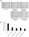
The effect of Aspergillus extract and aspochalasine A on HUVECs adhesion. HUVECs were treated with test samples (Aspergillus extract and aspochalasine A) or the DMSO vehicle (control) for 30 min. Cells that underwent adhesion were (A) imaged and (B) counted. Data are expressed as mean ± SEM. Similar results were obtained in three independent experiments; * p < 0.05 compared to the control value.
Figure 3
Figure 3
The effect of Aspergillus extract and aspochalasine A on HUVECs migration. (A) Representative images of HUVEC migration. (B) HUVECs were seeded into each well and treated with Aspergillus extract, aspochalasine A, or the DMSO vehicle (control) for 24 h. Cell migration was imaged and counted. Data are expressed as mean ± SEM. Similar results were obtained in three independent experiments; * p < 0.05 compared to the control value.
Figure 4
Figure 4
The effect of Aspergillus extract and aspochalasine A on HUVEC tube formation. (A) Representative images for tubule formation after treatment with the indicated concentrations of Aspergillus extract and aspochalasine A. (B) The length of the tubes was measured using the ImageJ software, and is represented as the percentage of tubule formation compared to the control. Data are expressed as mean ± SEM. Similar results were obtained in three independent experiments; * p < 0.05 compared to the control value.
Figure 5
Figure 5
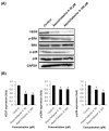
The effect of Aspergillus extract on angiogenic protein expression in HUVECs. (A) Western blot showing the levels of VEGF (21 kDa), phosphorylated ERK (42/44 kDa), ERK (42/44 kDa), phosphorylated p38 (38 kDa), and p38 (38 kDa) in HUVECs treated with Aspergillus extract at different concentrations for 24 h. (B) Graphs indicating quantification of the effect of Aspergillus extract on the angiogenic protein expression in HUVECs. Data are expressed as mean ± SEM. Similar results were obtained in three independent experiments; * p < 0.05 compared to the control value.
Figure 6
Figure 6
The effect of aspochalasine A on angiogenic protein expression in HUVECs. (A) Western blot indicating the levels of VEGF (21 kDa), phosphorylated ERK (42/44 kDa), ERK (42/44 kDa), phosphorylated p38 (38 kDa), and p38 (38 kDa) in HUVECs treated with aspochalasine A at different concentrations for 24 h. (B) Graphs representing quantification of effect of aspochalasine A on the angiogenic protein expressions in HUVECs. Data are expressed as mean ± SEM. Similar results were obtained in three independent experiments; * p < 0.05 compared to the control value.
Figure 7
Figure 7
The effect of Aspergillus extract and aspochalasine A on VEGFR-2 and PI3K/Akt signaling in HUVECs. (A,C) Western blot indicating the levels of phosphorylated VEGFR-2 (230 kDa), VEGFR-2 (210/ 230 kDa), phosphorylated Akt (60 kDa), and phosphorylated PI3K (85 kDa) in HUVECs treated with Aspergillus extract and aspochalasine A at different concentrations for 24 h. (B,D) Graphs representing quantification of effect of Aspergillus extract and aspochalasine A on the angiogenic protein expressions in HUVECs. Data are expressed as mean ± SEM. Similar results were obtained in three independent experiments; * p < 0.05 compared to the control value.
Figure 7
Figure 7
The effect of Aspergillus extract and aspochalasine A on VEGFR-2 and PI3K/Akt signaling in HUVECs. (A,C) Western blot indicating the levels of phosphorylated VEGFR-2 (230 kDa), VEGFR-2 (210/ 230 kDa), phosphorylated Akt (60 kDa), and phosphorylated PI3K (85 kDa) in HUVECs treated with Aspergillus extract and aspochalasine A at different concentrations for 24 h. (B,D) Graphs representing quantification of effect of Aspergillus extract and aspochalasine A on the angiogenic protein expressions in HUVECs. Data are expressed as mean ± SEM. Similar results were obtained in three independent experiments; * p < 0.05 compared to the control value.
Figure 8
Figure 8
Effect of Aspergillus extract and aspochalasine A on VEGF and VEGFR-2 mRNA expressions in HUVECs. Total RNA was extracted from the Aspergillus extract and aspochalasine A-treated HUVECs, and human VEGF-A mRNA or human VEGFR-2 mRNA expression were analyzed by reverse transcriptase (RT)-PCR. GAPDH was used as an internal control. Data are expressed as means ± SEM. Similar results were obtained from three independent experiments.
Figure 9
Figure 9
The in vivo anti-angiogenic effects of asperchalasine A and Aspergillus extract. (A) Images show representative blood vessel formation on the chorioallantoic membrane (CAM) following treatment with 10 and 20 μM asperchalasine A or 10 and 20 μg/mL Aspergillus extract for 48 h. (B) Calculations were based on the ratio of eggs with inhibited neovascularization relative to the total number of live eggs.

References

    1. Folkman J., Cotran R. Relation of vascular proliferation to tumor growth. Int. Rev. Exp. Pathol. 1976;16:207–248. - PubMed
    1. Folkman J. Angiogenesis in cancer, vascular, rheumatoid and other disease. Nat. Med. 1995;1:27–31. doi: 10.1038/nm0195-27. - DOI - PubMed
    1. Fukumura D., Gohongi T., Kadambi A., Izumi Y., Ang J., Yun C.O., Buerk D.G., Huang P.L., Jain R.K. Predominant role of endothelial nitric oxide synthase in vascular endothelial growth factor-induced angiogenesis and vascular permeability. Proc. Natl. Acad. Sci. USA. 2001;98:2604–2609. doi: 10.1073/pnas.041359198. - DOI - PMC - PubMed
    1. Park E.H., Park J.Y., Yoo H.S., Yoo J.E., Lee H.L. Assessment of the anti-metastatic properties of sanguiin H-6 in HUVECs and MDA-MB-231 human breast cancer cells. Bioorg. Med. Chem. Lett. 2016;26:3291–3294. doi: 10.1016/j.bmcl.2016.05.050. - DOI - PubMed
    1. Weidner N., Carroll P.R., Flax J., Blumenfeld W., Folkman J. Tumor angiogenesis correlates with metastasis in invasive prostate carcinoma. Am. J. Pathol. 1993;143:401–409. - PMC - PubMed

Publication types

MeSH terms

LinkOut - more resources