. 2019 Aug 12;20(16):3922.
doi: 10.3390/ijms20163922.

Impact of Astrocyte Depletion upon Inflammation and Demyelination in a Murine Animal Model of Multiple Sclerosis

Affiliations

Impact of Astrocyte Depletion upon Inflammation and Demyelination in a Murine Animal Model of Multiple Sclerosis

Lisa Allnoch et al. Int J Mol Sci. .

Abstract

Astrocytes play a key role in demyelinating diseases, like multiple sclerosis (MS), although many of their functions remain unknown. The aim of this study was to investigate the impact of astrocyte depletion upon de- and remyelination, inflammation, axonal damage, and virus distribution in Theiler`s murine encephalomyelitis (TME). Groups of two to six glial fibrillary acidic protein (GFAP)-thymidine-kinase transgenic SJL mice and SJL wildtype mice were infected with TME virus (TMEV) or mock (vehicle only). Astrocyte depletion was induced by the intraperitoneal administration of ganciclovir during the early and late phase of TME. The animals were clinically investigated while using a scoring system and a rotarod performance test. Necropsies were performed at 46 and 77 days post infection. Cervical and thoracic spinal cord segments were investigated using hematoxylin and eosin (H&E), luxol fast blue-cresyl violet (LFB), immunohistochemistry targeting Amigo2, aquaporin 4, CD3, CD34, GFAP, ionized calcium-binding adapter molecule 1 (Iba1), myelin basic protein (MBP), non-phosphorylated neurofilaments (np-NF), periaxin, S100A10, TMEV, and immunoelectron microscopy. The astrocyte depleted mice showed a deterioration of clinical signs, a downregulation and disorganization of aquaporin 4 in perivascular astrocytes accompanied by vascular leakage. Furthermore, astrocyte depleted mice showed reduced inflammation and lower numbers of TMEV positive cells in the spinal cord. The present study indicates that astrocyte depletion in virus triggered CNS diseases contributes to a deterioration of clinical signs that are mediated by a dysfunction of perivascular astrocytes.

Keywords: GFAP-thymidine kinase transgenic SJL mice; Theiler`s murine encephalomyelitis; aquaporin 4; astrocytes; blood-spinal cord barrier; glial fibrillary acidic protein.


Conflict of interest statement

The authors declare no conflict of interest.

Figures

Figure 1
Figure 1
Clinical investigation using a scoring system and a rotarod test. Astrocyte depletion between 28–46 days post infection (dpi; early phase) resulted in a deterioration of clinical signs at 45 and 46 dpi (A) and rotarod performance at 46 dpi (B) in Theiler’s murine encephalomyelitis virus (TMEV) infected, ganciclovir treated Gfap-transgenic (GSTG) mice as compared with TMEV infected, ganciclovir treated wildtype mice (WSTG). Astrocyte depletion between 56–77 dpi (late phase) resulted in a deterioration of clinical signs starting at 72 dpi (C) and rotarod performance at 7 and 77 dpi (D) in GSTG as compared with WSTG mice. Clinical and rotarod data are shown as median ± standard deviation. Significant differences between GSTG and the other groups, as obtained by the Kruskal–Wallis test, followed by Mann–Whitney U post hoc tests were indicated by , p < 0.05.
Figure 2
Figure 2
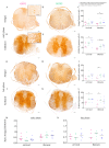
Astrocyte distribution during Theiler’s murine encephalomyelitis (TME). Immunohistochemistry targeting glial fibrillary acidic protein (GFAP) was used for the detection of astrocytes. At 46 days post infection (dpi; early phase) TMEV infected, ganciclovir treated, Gfap-transgenic mice showed a significantly reduced GFAP-positive area in the white matter of the thoracic spinal cord (A,C) compared to TMEV infected, ganciclovir treated wildtype (WSTG; B,D) and TMEV infected, natrium chloride treated wildtype (WSTP; E) mice. At 77 dpi (late phase) TMEV infected, ganciclovir treated, Gfap-transgenic mice showed a significantly reduced GFAP-positive area in the white matter of the cervical and thoracic spinal cord segment (F,H) compared with WSTG (G,I) and WSTP (J). Insert shows the morphology of an intralesional astrocyte in a GSTG animal (H). Data are shown as scatter dot plots. Significant differences between the groups obtained by Kruskal-Wallis test followed by Mann–Whitney U post hoc tests were indicated by *, p < 0.05. Bars represent 100 µm in overviews and 50 µm in the insert.
Figure 3
Figure 3
Astrocyte phenotype identification and quantification. Identification and quantification of astrocyte phenotypes in the ventral part of the spinal cord white matter were performed using immunohistochemistry identifying A1 (Amigo2, insert in A) and A2 (S100A10, insert in D). Astrocyte depletion resulted in a reduced number of Amigo2 positive cells in the thoracic spinal cord of TMEV infected, ganciclovir treated, Gfap-transgenic mice (GSTG) mice at 46 days post infection (dpi; AC) and 77 dpi (late phase; G–I). Immunohistochemistry for S100A10 (A2 astrocytes) detected a significant reduction of the A2 subpopulation in the cervical and thoracic spinal cord at 46 dpi (early phase; DF) and 77 dpi (late phase; JL). Statistical analysis did not reveal a significant difference between the ratios of A1 and A2 astrocytes in all investigated groups. However, at 46 dpi (M) and 77 dpi (N) a variable predominance of A2 astrocytes in all groups was observed. Data are shown as scatter dot plots. Significant differences between the groups obtained by Kruskal–Wallis test followed by Mann–Whitney U post hoc tests were indicated by *, p < 0.05. Bars represent 100 µm in overviews and 50 µm in the inserts.
Figure 4
Figure 4
Impact of astrocyte depletion upon aquaporin 4 (AQP4) expression. Immunohistochemistry targeting AQP4 revealed a significant reduction of AQP4 positive area in the cervical and thoracic spinal cord segment of TMEV infected, ganciclovir treated, Gfap-transgenic mice (GSTG) mice at 46 days post infection (dpi; early phase; AE). At 77 dpi (late phase), GSTG mice showed a significant reduction of AQP4 positive area in the thoracic spinal cord (FJ). Data are shown as scatter dot plots. Significant differences between the groups obtained by Kruskal-Wallis test followed by Mann–Whitney U post hoc tests were indicated by *, p < 0.05. Bars represent 100 µm.
Figure 5
Figure 5
Immunoelectron microscopy targeting aquaporin 4 (AQP4). Immunoelectron microscopy of the thoracic spinal cord segment was performed for the localization of AQP4 within perivascular astrocytes. A reduced amount and an irregular distribution of AQP4 protein along the cytoplasmic membrane of perivascular astrocyte end-feet (arrows in B) in TMEV infected, ganciclovir treated, Gfap-transgenic (GSTG, A,B) mice compared to control mice (C,D) at 77 days post infection was detected. The distribution of AQP4 along the cytoplasmic membrane is shown in higher magnification (inserts in A,C). The reduced expression of AQP4 in GSTG animals was associated with a shortening and disorganization of intermediate filaments (asterisks in A,B) in comparison to control animals (arrowheads in C,D). Magnifications are 40,000× in A, 25,000× in B and 31,500 in C,D.
Figure 6
Figure 6
Detection of vascular leakage. Immunohistochemistry targeting CD34 was used for the detection of vascular leakage within the spinal cord white matter. Variable CD34 labeling was predominantly observed in the ventral part of the cervical and thoracic spinal cord white matter at 46 (early phase; A,B) and 77 (late phase; C,D) days post infection in all groups. CD34 labeling is shown in higher magnification (insert in C). Bars represent 100 µm in the overviews and 50 µm in the insert.
Figure 7
Figure 7
Quantification of meningeal and perivascular inflammation. Meningitis and perivascular inflammation in the cervical and thoracic spinal cord white matter were quantified using hematoxylin and eosin (H&E) staining. Statistical analysis revealed a significant reduction of meningitis in TMEV infected, ganciclovir treated, Gfap-transgenic (GSTG) mice at 77 days post infection (dpi; late phase; AC). Perivascular inflammation in the white matter of the cervical spinal cord at 46 dpi (early phase; E) and of the thoracic spinal cord at 77 dpi (late phase; F) was significantly reduced in GSTG animals. The inserts in A and D show higher magnifications. Data are shown as scatter dot plots. Significant differences between the groups obtained by Kruskal–Wallis test followed by Mann–Whitney U post hoc tests were indicated by *, p < 0.05. Bars represent 100 µm in the overviews and 50 µm in the inserts.
Figure 8
Figure 8
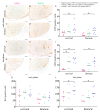
Immunophenotyping and quantification of inflammatory cells. T lymphocytes (CD3) were quantified in the cervical and thoracic spinal cord using immunohistochemistry. TMEV infected, ganciclovir treated, Gfap-transgenic (GSTG) mice showed a reduced number of CD3 positive cells in the cervical and thoracic spinal cord at 46 days post infection (dpi; early phase; AE) and 77 dpi (late phase; FJ). Immunohistochemistry targeting ionized calcium-binding adapter molecule 1 (Iba1) was used for the quantification of microglia/macrophages. While at 46 dpi no significant differences between the groups were detected (K) at 77 dpi the number of Iba1 labeled cells in the cervical and thoracic spinal cord was significantly reduced in GSTG mice (L). Data are shown as scatter dot plots. Significant differences between the groups obtained by Kruskal–Wallis test followed by Mann–Whitney U post hoc tests were indicated by *, p < 0.05. Bars represent 100 µm.
Figure 9
Figure 9
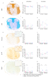
Quantification of demyelination, Schwann cell remyelination, axonal damage, and virus protein. Myelin basic protein (MBP; A–C) immunohistochemistry and luxol fast blue-cresyl violet (LFB; DF) staining were used to quantify demyelination after astrocyte depletion. Accentuated in the ventral parts of the spinal cord white matter demyelination occurred in a variable degree in all groups. However, statistical analysis did not reveal a significant difference between TMEV infected, ganciclovir treated, Gfap-transgenic (GSTG) mice and the other groups (B,C,E,F). Periaxin immunohistochemistry was used to detect Schwann cell remyelination (GI). At 77 days post infection (dpi; late phase) the cervical and thoracic spinal cords showed low numbers of Schwann cells in all groups (I). However, statistical analysis did not reveal a significant difference of periaxin positive Schwann cell numbers in GSTG mice compared to the control groups (H,I). Immunohistochemistry detecting non-phosphorylated-neurofilaments (np-NF) was used to detect and quantify axonal damage (JL). Accentuated in the ventral parts of the white matter variable numbers of np-NF labeled axons were detected in all groups. Statistical analysis did not reveal a significant difference of axonal damage in GSTG mice compared to the controls at 46 dpi (early phase; K) and 77 dpi (late phase; L). Virus protein was detected and quantified by Theiler’s murine encephalomyelitis virus (TMEV) immunohistochemistry (MO). Virus positive cells were present in the cervical and thoracic spinal cord of all groups. GSTG mice revealed a significantly reduced number of TMEV positive cells in the cervical spinal cord at 46 dpi (early phase, N) and in the cervical and thoracic spinal cord at 77 dpi (late phase; O). The data are shown as scatter dot plots. Significant differences between the groups obtained by Kruskal-Wallis test followed by Mann–Whitney U post hoc tests were indicated by *, p < 0.05. Bars represent 100 µm in the overviews and 50 µm in the inserts.
Figure 10
Figure 10
Presumed roles of astrocytes in multiple sclerosis (MS) and Theiler’s murine encephalomyelitis. Functions of astrocytes in MS are shown on the left side of the scheme [8,48,49,50,51]. They include an interaction with neurons by secreting neurotrophic as well as neurotoxic factors. Microglia/macrophage recruitment and T lymphocyte extravasation are induced by astrocytes as well. Furthermore, astrocytes are able to inhibit oligodendrocyte precursor cell (OPC) migration and induce oligodendrocyte death. Secretion of matrix metalloproteinases (MMPs) like MMP-9 significantly contributes to an impairment of the blood brain barrier (BBB). The right side of the schematic drawing summarizes the effects of astrocyte depletion in Theiler’s murine encephalomyelitis (TME). Astrocyte depletion during the early and late phase of TME resulted in a lower number of TMEV positive cells and a reduction of spinal cord inflammation with no effect on de- and remyelination. Furthermore, astrocyte depletion was associated with a reduction and an irregular arrangement of aquaporin 4 (AQP4) water channels, as well as a shortening and disorganization of intermediate filaments in perivascular astrocytes. BDNF, Brain-derived neurotrophic factor; CCL, chemokine ligand; GDNF, Glial cell line-derived neurotrophic factor; NO, nitric oxide; NT-3, Neurotrophin-3; TNF, tumor necrosis factor.
Figure 11
Figure 11
Study design. Groups of 2–6 GFAP-thymidine-kinase transgenic SJL mice and SJL wildtype mice were infected with Theiler’s murine encephalomyelitis virus (TMEV) or mock (vehicle only). Astrocyte depletion was induced by once daily intraperitoneal administration of ganciclovir from 28 to 46 (early phase) or 56 to 77 (late phase) days post infection. Red line indicates the ganciclovir/NaCl treatment period.

References

    1. Sofroniew M.V., Vinters H.V. Astrocytes: Biology and pathology. Acta Neuropathol. 2010;119:7–35. doi: 10.1007/s00401-009-0619-8. - DOI - PMC - PubMed
    1. Ludwin S.K., Rao V., Moore C.S., Antel J.P. Astrocytes in multiple sclerosis. Mult. Scler. 2016;22:1114–1124. doi: 10.1177/1352458516643396. - DOI - PubMed
    1. Liddelow S.A., Barres B.A. Reactive astrocytes: Production, function, and therapeutic potential. Immunity. 2017;46:957–967. doi: 10.1016/j.immuni.2017.06.006. - DOI - PubMed
    1. Jäkel S., Dimou L. Glial cells and their function in the adult brain: A journey through the history of their ablation. Front. Cell. Neurosci. 2017;11:24. doi: 10.3389/fncel.2017.00024. - DOI - PMC - PubMed
    1. Nash B., Ioannidou K., Barnett S.C. Astrocyte phenotypes and their relationship to myelination. J. Anat. 2011;219:44–52. doi: 10.1111/j.1469-7580.2010.01330.x. - DOI - PMC - PubMed

MeSH terms