

Cellular and Molecular Mechanisms in the Pathogenesis of Classical, Vascular, and Hypermobile Ehlers‒Danlos Syndromes
- PMID: 31409039
- PMCID: PMC6723307
- DOI: 10.3390/genes10080609
Cellular and Molecular Mechanisms in the Pathogenesis of Classical, Vascular, and Hypermobile Ehlers‒Danlos Syndromes
Abstract
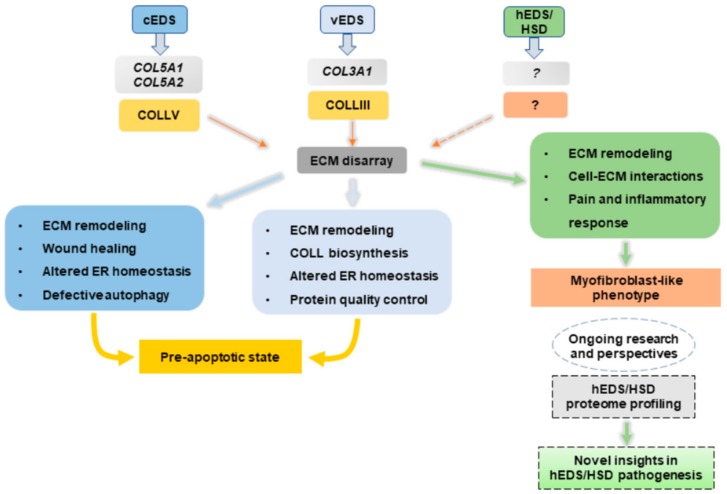
The Ehlers‒Danlos syndromes (EDS) constitute a heterogenous group of connective tissue disorders characterized by joint hypermobility, skin abnormalities, and vascular fragility. The latest nosology recognizes 13 types caused by pathogenic variants in genes encoding collagens and other molecules involved in collagen processing and extracellular matrix (ECM) biology. Classical (cEDS), vascular (vEDS), and hypermobile (hEDS) EDS are the most frequent types. cEDS and vEDS are caused respectively by defects in collagen V and collagen III, whereas the molecular basis of hEDS is unknown. For these disorders, the molecular pathology remains poorly studied. Herein, we review, expand, and compare our previous transcriptome and protein studies on dermal fibroblasts from cEDS, vEDS, and hEDS patients, offering insights and perspectives in their molecular mechanisms. These cells, though sharing a pathological ECM remodeling, show differences in the underlying pathomechanisms. In cEDS and vEDS fibroblasts, key processes such as collagen biosynthesis/processing, protein folding quality control, endoplasmic reticulum homeostasis, autophagy, and wound healing are perturbed. In hEDS cells, gene expression changes related to cell-matrix interactions, inflammatory/pain responses, and acquisition of an in vitro pro-inflammatory myofibroblast-like phenotype may contribute to the complex pathogenesis of the disorder. Finally, emerging findings from miRNA profiling of hEDS fibroblasts are discussed to add some novel biological aspects about hEDS etiopathogenesis.
Keywords: Ehlers‒Danlos syndrome; autophagy; collagen III; collagen V; endoplasmic reticulum; extracellular matrix; fibroblast-to-myofibroblast transition; miRNA; transcriptome; wound healing.
Conflict of interest statement
The authors declare no conflicts of interests.
Figures
References
Publication types
MeSH terms
LinkOut - more resources
Full Text Sources
Medical
