. 2019 Aug 13;17(1):265.
doi: 10.1186/s12967-019-2013-1.

Renal temperature reduction progressively favors mitochondrial ROS production over respiration in hypothermic kidney preservation

Affiliations

Renal temperature reduction progressively favors mitochondrial ROS production over respiration in hypothermic kidney preservation

Koen D W Hendriks et al. J Transl Med. .

Abstract

Background: Hypothermia, leading to mitochondrial inhibition, is widely used to reduce ischemic injury during kidney preservation. However, the exact effect of hypothermic kidney preservation on mitochondrial function remains unclear.

Methods: We evaluated mitochondrial function [i.e. oxygen consumption and production of reactive oxygen species (ROS)] in different models (porcine kidney perfusion, isolated kidney mitochondria, and HEK293 cells) at temperatures ranging 7-37 °C.

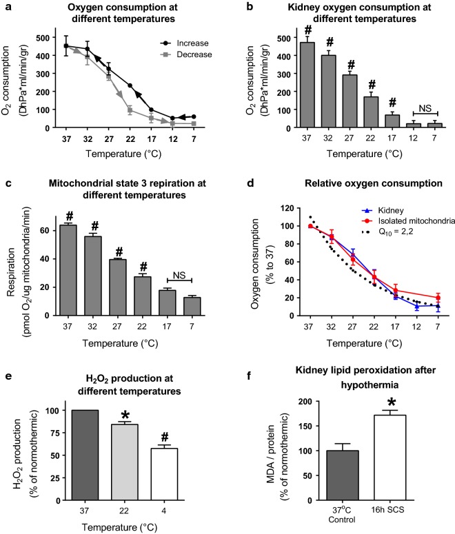
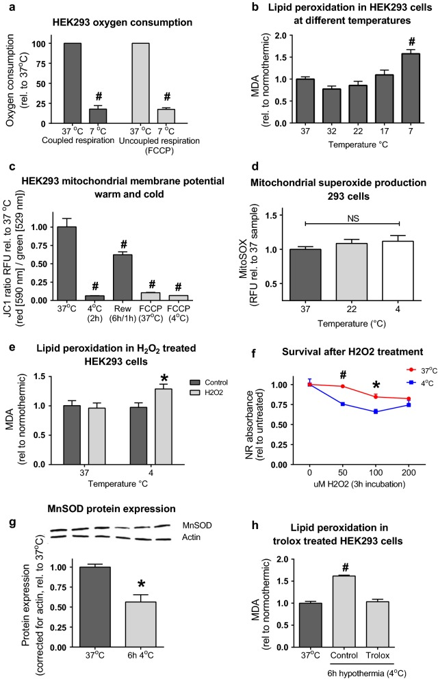
Results: Lowering temperature in perfused kidneys and isolated mitochondria resulted in a rapid decrease in oxygen consumption (65% at 27 °C versus 20% at 7 °C compared to normothermic). Decreased oxygen consumption at lower temperatures was accompanied by a reduction in mitochondrial ROS production, albeit markedly less pronounced and amounting only 50% of normothermic values at 7 °C. Consequently, malondialdehyde (a marker of ROS-induced lipid peroxidation) accumulated in cold stored kidneys. Similarly, low temperature incubation of kidney cells increased lipid peroxidation, which is due to a loss of ROS scavenging in the cold.

Conclusions: Lowering of temperature highly affects mitochondrial function, resulting in a progressive discrepancy between the lowering of mitochondrial respiration and their production of ROS, explaining the deleterious effects of hypothermia in transplantation procedures. These results highlight the necessity to develop novel strategies to decrease the formation of ROS during hypothermic organ preservation.

Keywords: Hypothermic preservation; Kidney transplantation; Machine perfusion; Mitochondrial function; Reactive oxygen species.


Conflict of interest statement

The authors declare that they have no competing interests.

Figures

Fig. 1
Fig. 1
Temperature effects on porcine kidneys. a Oxygen consumption [expressed as ΔhPa (ml/min/gr)] in perfused porcine kidneys. The increasing and decreasing curve are expressed. Based on four independent experiments, expressed as mean, error bars represent SEM. b Average of whole kidney oxygen consumption per temperature. Expressed as mean, error bars represent SEM. #Significant (ANOVA with Bonferroni posthoc, p < 0.001). c State three respiration in isolated mitochondria, in response to glutamate and malate with and an ADP generating system. Based on five independent experiments, expressed as mean, error bars represent SEM. #Significant (ANOVA with Bonferroni posthoc, p < 0.001). d Oxygen consumption relative to 37 °C for perfused kidneys and isolated kidney mitochondria. The theoretical Q10 line is plotted (Q10 = 2). e Relative ROS production, measured as mitochondrial H2O2 production at different temperatures in isolated mitochondria from porcine kidneys. N = 4, expressed as percentage of its normothermic control, error bars represent SEM. *p < 0.01, #p < 0.001 (ANOVA with Bonferroni posthoc). f ROS damage, measured as lipid peroxidation in porcine kidneys before and after 16 h of static cold storage in UW at 4 °C. N = 4, expressed as relative to normothermic, error bars represent SEM. *p < 0.01, (Students t test)
Fig. 2
Fig. 2
Temperature effects on HEK293 cells. a State three oxygen consumption at normothermic (37 °C) and hypothermic (4 °C) circumstances in coupled and uncoupled HEK293 cells. Based on three independent experiments, expressed as rel. to normothermic, error bars represent SEM. #p < 0.001 (Students T test). b Lipid peroxidation (MDA) at different temperatures in HEK293 cells. N = 6, expressed as MDA levels rel. to normothermic, error bars represent SEM. #p < 0.001 compared to 37 °C (ANOVA with Bonferroni posthoc). c Mitochondrial membrane potential in normothermic, hypothermic and rewarmed (rew) HEK293 cells. N = 6, expressed as JC1 ratio RFU rel. to 37 °C (red [590 nm]/green [529 nm]), error bars represent SEM. #p < 0.001 compared to 37 °C (ANOVA with Bonferroni posthoc). FCCP (carbonyl cyanide 4-(trifluoromethoxy)phenylhydrazone 1 μM) as uncoupled control. d Mitochondrial superoxide production in HEK293 cells, incubated for 90 min at 37°, 22° or 4°. N = 6, expressed as mitosox RFU, error bars represent SEM. #p < 0.001 compared to 37 °C (ANOVA with Bonferroni posthoc). e Lipid peroxidation in H2O2 stimulated normothermic and hypothermic treated HEK293 cells. N = 6, expressed as MDA levels rel. to normothermic, error bars represent SEM. *p < 0.05 compared to control (Student t test). f HEK293 survival after 3 h H2O2 exposure at different concentrations and temperatures. N = 3, expressed as Neutral Red absorbance rel. to untreated control. *p < 0.05 compared to non-treated (Student t test), #p < 0.001 (Students T test). g MnSOD protein expression in normothermic and 6 h hypothermic (4 °C) HEK293 cells. Expressed as corrected values for actin, rel. to 37 °C, error bars represent SEM. *p < 0.05 (Student t test). Full membranes shown in Additional file 1: Figure S3B. h MDA levels in trolox treated 6 h hypothermic HEK293 cells. N = 3, expressed as MDA levels rel. to normothermic, error bars represent SEM. #p < 0.001 compared to 37 °C (ANOVA with Bonferroni posthoc)

References

    1. Pezzati D, Ghinolfi D, De Simone P, Balzano E, Filipponi F. Strategies to optimize the use of marginal donors in liver transplantation. World J Hepatol. 2015;7(26):2636–2647. doi: 10.4254/wjh.v7.i26.2636. - DOI - PMC - PubMed
    1. Attia M, Silva MA, Mirza DF. The marginal liver donor—an update. Transpl Int. 2008;21(8):713–724. doi: 10.1111/j.1432-2277.2008.00696.x. - DOI - PubMed
    1. Saidi RF, Kenari SKH. Liver ischemia/reperfusion injury: an overview. J Invest Surg. 2014;27(6):366–379. doi: 10.3109/08941939.2014.932473. - DOI - PubMed
    1. Chouchani ET, Pell VR, Gaude E, Aksentijević D, Sundier SY, Robb EL, et al. Ischaemic accumulation of succinate controls reperfusion injury through mitochondrial ROS. Nature. 2014;515(7527):431–435. doi: 10.1038/nature13909. - DOI - PMC - PubMed
    1. Schlegel A, Kron P, Dutkowski P. Hypothermic oxygenated liver perfusion: basic mechanisms and clinical application. Curr Transplant Rep. 2015;2(1):52–62. doi: 10.1007/s40472-014-0046-1. - DOI - PMC - PubMed

Publication types