. 2019 Nov 6;27(11):1992-2004.
doi: 10.1016/j.ymthe.2019.07.016. Epub 2019 Jul 30.

Pharmacological and Genetic Inhibition of Caveolin-1 Promotes Epithelialization and Wound Closure

Affiliations

Pharmacological and Genetic Inhibition of Caveolin-1 Promotes Epithelialization and Wound Closure

Ivan Jozic et al. Mol Ther. .

Abstract

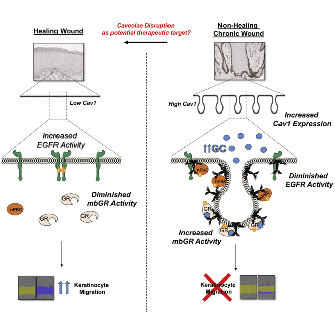
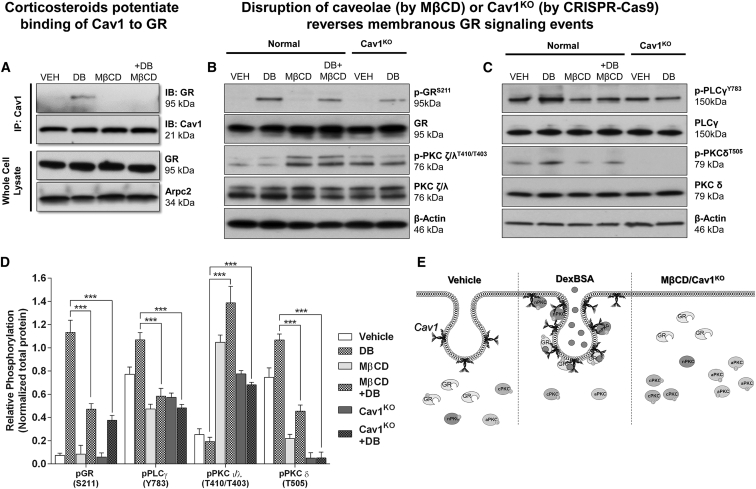
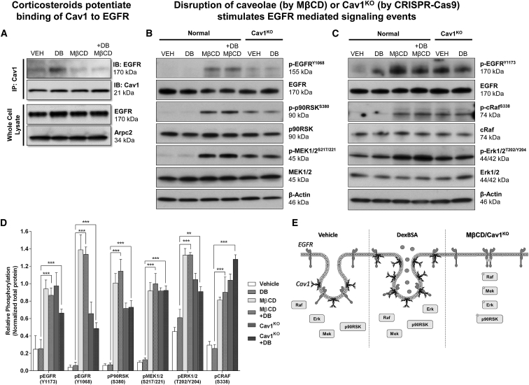
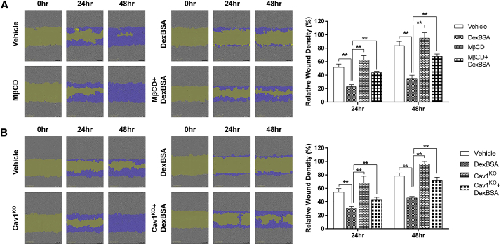
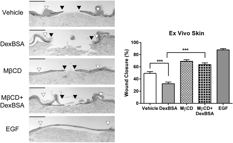
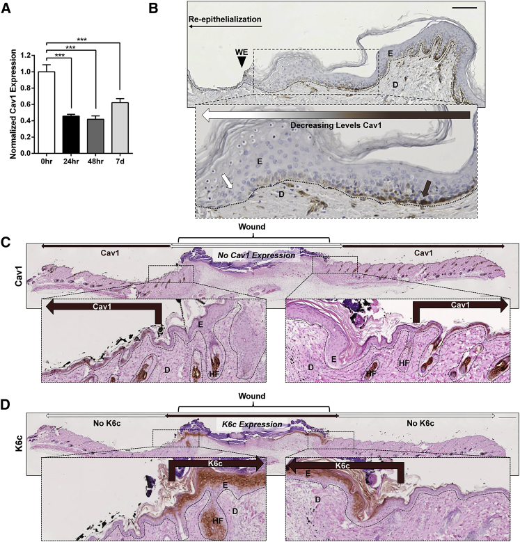
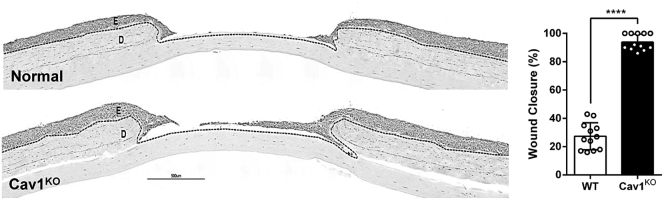
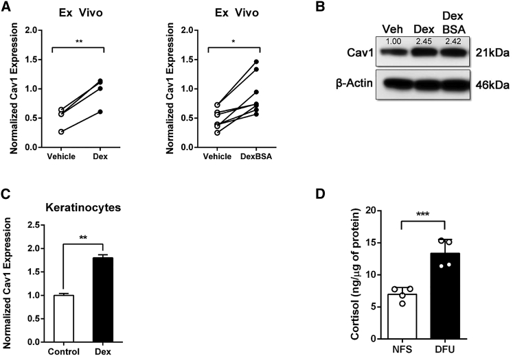
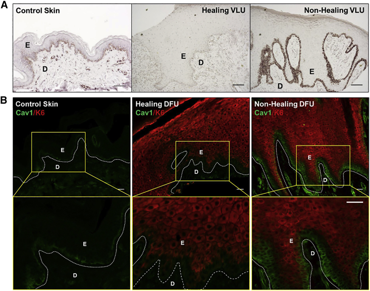
Chronic wounds-including diabetic foot ulcers, venous leg ulcers, and pressure ulcers-represent a major health problem that demands an urgent solution and new therapies. Despite major burden to patients, health care professionals, and health care systems worldwide, there are no efficacious therapies approved for treatment of chronic wounds. One of the major obstacles in achieving wound closure in patients is the lack of epithelial migration. Here, we used multiple pre-clinical wound models to show that Caveolin-1 (Cav1) impedes healing and that targeting Cav1 accelerates wound closure. We found that Cav1 expression is significantly upregulated in wound edge biopsies of patients with non-healing wounds, confirming its healing-inhibitory role. Conversely, Cav1 was absent from the migrating epithelium and is downregulated in acutely healing wounds. Specifically, Cav1 interacted with membranous glucocorticoid receptor (mbGR) and epidermal growth factor receptor (EGFR) in a glucocorticoid-dependent manner to inhibit cutaneous healing. However, pharmacological disruption of caveolae by MβCD or CRISPR/Cas9-mediated Cav1 knockdown resulted in disruption of Cav1-mbGR and Cav1-EGFR complexes and promoted epithelialization and wound healing. Our data reveal a novel mechanism of inhibition of epithelialization and wound closure, providing a rationale for pharmacological targeting of Cav1 as potential therapy for patients with non-healing chronic wounds.

Keywords: Caveolin-1; chronic wounds; diabetic foot ulcers; skin re-epithelialization; wound healing.


Figures

None
Graphical abstract
Figure 1
Figure 1
Cav-1 Binds to mbGR, and Disruption of Caveolae Results in a Reversal of mbGR Effects in Primary Human Keratinocytes (A) Primary human keratinocytes were stimulated with vehicle (DMSO) or 100 nM Dex-BSA (hereinafter referred to as DB) for 30 min in the presence or absence of 2% MβCD, followed by Cav1 co-immunoprecipitation. (B and C) Levels of total GR, phospho-GRS211, atypical total PKC ζ/λ, and phospho-PKC ζ/λT410/T403 (B) and levels of total PLCγ, p-PLCγY783, novel total PKC δ, and phospho-PKC δT505 (C) were assessed by immunoblotting; Arpc2 and β-actin were used as loading controls. Whole-cell lysate membranes were stripped and re-probed with appropriate antibodies. (D) Signals were quantified as amount of phosphorylated protein over total protein. Experiments were done in triplicates, with error bars corresponding to SDs from 3 biological triplicates with at least three technical replicates. ***p ≤ 0.001, two-way ANOVA. (E) Treatment with DexBSA causes interaction of Cav1 with pGR and nPKC and potentiates mbGR-mediated signaling events, which can be ameliorated either by disrupting caveolae (by MβCD) or knocking out Cav1 from the cells (Cav1KO).
Figure 2
Figure 2
Cav-1 Binds Also to EGFR, and Disruption of Caveolae Results in a Rescue of EGFR Signaling in Primary Human Keratinocytes (A) Primary human keratinocytes were stimulated with vehicle (DMSO) or 100 nM Dex-BSA for 2 min in the presence or absence of 2% MβCD, followed by Cav1 co-immunoprecipitation. (B and C) Levels of total EGFR, p-EGFRY1068, total cRaf, p-cRafS338, total Erk1/2, p-Erk1/2T202/Y204 (B) and levels of p-EGFRY1173, total p90RSK, p-p90RSKS380, total MEK1/2, and p-MEK1/2S217/221 (C) were assessed by immunoblotting. Whole-cell lysate membranes were stripped and re-probed with appropriate antibodies. (D) Signals were quantified as amount of phosphorylated protein over total protein. Experiments were done in triplicates, with error bars corresponding to SDs from biological triplicates with at least three technical replicates. **p ≤ 0.01; ***p ≤ 0.001, two-way ANOVA. (E) Treatment with DexBSA causes interaction of Cav1 with EGFR and inhibits downstream EGFR-mediated signaling events, which can be reversed either by disrupting caveolae (by MβCD) or knocking out Cav1 from the cells (Cav1KO).
Figure 3
Figure 3
Pharmacologic Disruption of Caveolae or Genetic Targeting of Cav1 Reverses GC-Mediated Inhibition of Keratinocyte Migration (A and B) Control (A) or Cav1-deficient (B) human keratinocytes were pre-treated with 4 μg/mL mitomycin-C in the presence or absence of 2% MβCD and either vehicle (DMSO) or 100 nM Dex-BSA. Cells were wounded by a scratch, and their migration was assessed at the time of the scratch (0 h) and every 2 h for 48 h. Keratinocyte migration was quantified by comparing relative wound density using the Cell Migration Analysis software module (Essen BioScience) 24 and 48 h after the initial scratches were made; purple corresponds to repopulation of the wound over time by migrating cells. Error bars correspond to SD from 3 independent experiments with 16 technical replicates each. **p ≤ 0.01, two-way ANOVA.
Figure 4
Figure 4
Disruption of Caveolae Accelerates Wound Healing and Rescues mbGR-Mediated Inhibition of Wound Closure in Human Skin Human skin was wounded and maintained at the air-liquid interface in the presence or absence of 2% (w/v) MβCD, 100 nM Dex-BSA, or 25 ng/mL EGF. Wound healing was assessed at day 5 post-wounding, a time point when exponential epithelialization occurs. Gross photos show visual signs of closure and correspond to the histology assessments. White arrowheads point to the initial site of wounding, while black arrowheads point to the wound edge of the migrating epithelial tongue. Scale bars, 500 μm. Graphs represent summarized migration rates; error bars correspond to SD from 18 biological samples from 6 independent experiments with 3 technical replicates. ***p < 0.001, two-way ANOVA.
Figure 5
Figure 5
Cav1 Expression Is Spatiotemporally Downregulated in Both Human Ex Vivo and Mouse In Vivo Wounds (A and B) Healthy human skin was wounded and maintained at the air-liquid interface for 0 h, 24 h, 48 h, 96 h, and 7 days prior to assessment of Cav1 expression by (A) qPCR and (B) immunohistochemistry from 15 biological specimens from 5 independent experiments with 3 technical replicates. ***p ≤ 0.001, Student t test. Brown arrows point to expression of Cav1 in normal skin away from the wound, while white arrows indicate absence of Cav1 at the wound edge of the migrating epithelial tongue ex vivo. Scale bar, 100 μm. (C) Cav1 expression assessed by immunohistochemistry in murine excisional wounds 6 days post-wounding. White double arrows indicate wound area, brown arrows point to expression of Cav1 in skin away from the wound, while white arrows indicate absence of Cav1 from the wound edge of the migrating epithelial tongue in vivo. Bottom panel represents enlargement of the wound edge area. (D) Cytokeratin 6c (K6c) was used as a marker for migrating keratinocytes of the newly formed epidermis in murine excisional wounds; bottom panel represents enlargement of the wound edge area; dashed lines demarcate epidermal-dermal boundary. E, epidermis; D, dermis WE, wound edge; HF, hair follicle; n = 6. Scale bar, 500 μm.
Figure 6
Figure 6
Cav1 Knockdown Accelerates Wound Closure in Human Skin Equivalents Control and Cav1KO organotypic skin was wounded, and wound closure was quantified by histomorphometric analyses; dashed lines demarcate epidermal-dermal boundary. Scale bar, 500 μm. Graph represents quantification of wound closure; ****p ≤ 0.0001, pairwise Student’s t test, 12 biological samples from 3 independent experiments with 4 technical replicates, two tailed. E, epidermis; D, dermis.
Figure 7
Figure 7
Induction of Cav1 Contributes to Inhibition of Healing in Human Skin (A and B) Ex vivo human wounds were treated daily with 1 μM dexamethasone (Dex) or Dex-BSA, after which relative expression of Cav1 was determined by (A) qRT-PCR (pairwise Student’s t test from 4 [for Dex-treated skin] or 8 [for Dex-BSA-treated skin] biological samples; *p ≤ 0.05 and **p ≤ 0.01, two tailed) and (B) western blotting. (C) Human keratinocyte HaCaT cells were treated with 1 μM Dex for 48 h, and levels of Cav1 were determined by qRT-PCR (t test from 3 independent experiments each consisting of 3 technical replicates; **p ≤ 0.01, two tailed). (D) Cortisol levels of 4 normal skin and 4 diabetic foot ulcer wounds were measured by ELISA normalized by micrograms of tissue (***p ≤ 0.001, t test, two tailed).
Figure 8
Figure 8
Cav1 Expression Is Upregulated in Chronic Wound Epidermis, and Its Levels Correlate with Clinical Outcomes of Healing (A and B) Location-matched control skin as well as healing/non-healing (A) VLU and (B) DFU skin were subjected to Cav1 peroxidase or immunofluorescence staining. Healing outcome of VLUs and DFUs was determined based on wound size monitored over 4 weeks., D, dermis; E, epidermis. Scale bars, 200 μm.

Similar articles

Cited by

References

    1. Eming S.A., Martin P., Tomic-Canic M. Wound repair and regeneration: mechanisms, signaling, and translation. Sci. Transl. Med. 2014;6:265sr6. - PMC - PubMed
    1. Pastar I., Stojadinovic O., Yin N.C., Ramirez H., Nusbaum A.G., Sawaya A., Patel S.B., Khalid L., Isseroff R.R., Tomic-Canic M. Epithelialization in wound healing: a comprehensive review. Adv. Wound Care (New Rochelle) 2014;3:445–464. - PMC - PubMed
    1. Pastar I., Wong L.L., Egger A.N., Tomic-Canic M. Descriptive vs mechanistic scientific approach to study wound healing and its inhibition: is there a value of translational research involving human subjects? Exp. Dermatol. 2018;27:551–562. - PMC - PubMed
    1. Stojadinovic O., Brem H., Vouthounis C., Lee B., Fallon J., Stallcup M., Merchant A., Galiano R.D., Tomic-Canic M. Molecular pathogenesis of chronic wounds: the role of beta-catenin and c-myc in the inhibition of epithelialization and wound healing. Am. J. Pathol. 2005;167:59–69. - PMC - PubMed
    1. Vukelic S., Stojadinovic O., Pastar I., Vouthounis C., Krzyzanowska A., Das S., Samuels H.H., Tomic-Canic M. Farnesyl pyrophosphate inhibits epithelialization and wound healing through the glucocorticoid receptor. J. Biol. Chem. 2010;285:1980–1988. - PMC - PubMed

Publication types

MeSH terms