. 2019 Aug 13;10(4):e01780-19.
doi: 10.1128/mBio.01780-19.

Identification of the Cell-Surface Protease ADAM9 as an Entry Factor for Encephalomyocarditis Virus

Affiliations

Identification of the Cell-Surface Protease ADAM9 as an Entry Factor for Encephalomyocarditis Virus

Jim Baggen et al. mBio. .

Abstract

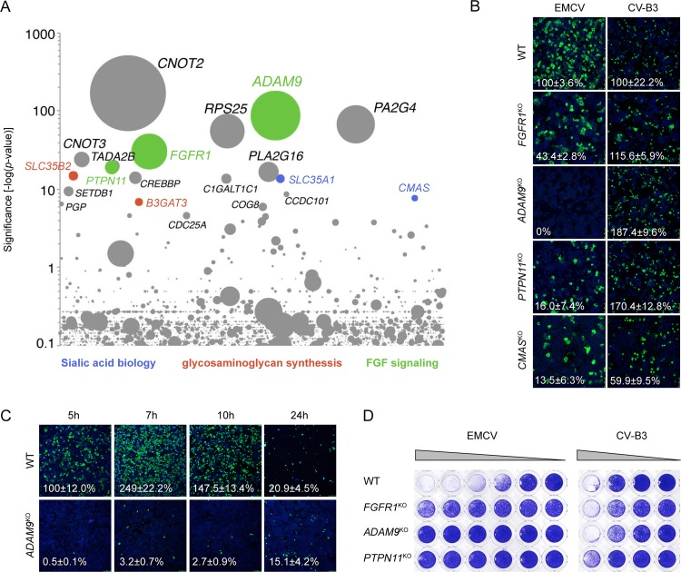
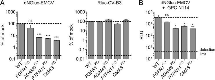
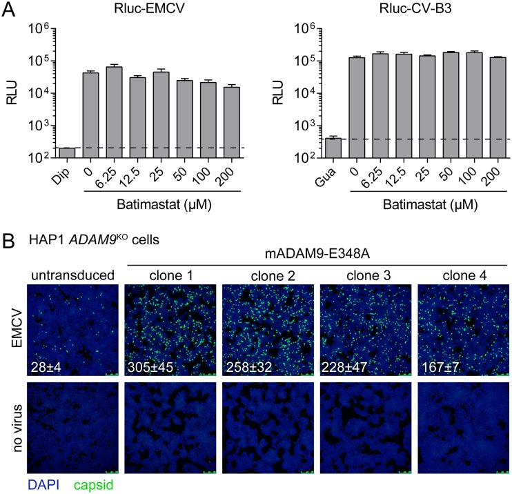
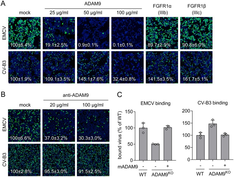
Encephalomyocarditis virus (EMCV) is an animal pathogen and an important model organism, whose receptor requirements are poorly understood. Here, we employed a genome-wide haploid genetic screen to identify novel EMCV host factors. In addition to the previously described picornavirus receptors sialic acid and glycosaminoglycans, this screen unveiled important new host factors for EMCV. These factors include components of the fibroblast growth factor (FGF) signaling pathway, such as the potential receptors FGFR1 and ADAM9, a cell-surface metalloproteinase. By employing various knockout cells, we confirmed the importance of the identified host factors for EMCV infection. The largest reduction in infection efficiency was observed in cells lacking ADAM9. Pharmacological inhibition of the metalloproteinase activity of ADAM9 did not affect virus infection. Moreover, reconstitution of inactive ADAM9 in knockout cells restored susceptibility to EMCV, pointing to a proteinase-independent role of ADAM9 in mediating EMCV infection. Using neutralization assays with ADAM9-specific antiserum and soluble receptor proteins, we provided evidence for a role of ADAM9 in EMCV entry. Finally, binding assays showed that ADAM9 facilitates attachment of EMCV to the cell surface. Together, our findings reveal a role for ADAM9 as a novel receptor or cofactor for EMCV.IMPORTANCE EMCV is an animal pathogen that causes acute viral infections, usually myocarditis or encephalitis. It is thought to circulate mainly among rodents, from which it is occasionally transmitted to other animal species, including humans. EMCV causes fatal outbreaks of myocarditis and encephalitis in pig farms and zoos, making it an important veterinary pathogen. Although EMCV has been widely used as a model to study mechanisms of viral disease in mice, little is known about its entry mechanism. Here, we employ a haploid genetic screen for EMCV host factors and identify an essential role for ADAM9 in EMCV entry.

Keywords: disintegrin and metalloproteinase domain-containing protein 9 (ADAM9); encephalomyocarditis virus; haploid genetic screen.


Figures

FIG 1
FIG 1
A genome-wide haploid screen identifies novel host factors for EMCV. (A) Bubble plot indicating the significance of enrichment of gene trap insertions in genes (y axis) in an EMCV-infected cell population compared to an uninfected control population. Each bubble represents a gene, with size corresponding to the number of gene trap insertions per gene. Genes were randomly distributed on the x axis. The names of the top 20 hits are indicated. (B) HAP1 cells deficient for various genes were infected with EMCV or coxsackievirus and adenovirus receptor (CAR)-binding CV-B3, followed by staining of capsid proteins (EMCV) or 3A protein (CV-B3) and nuclei (blue) at 5 h postinfection. Representative confocal micrographs are shown. The values (percentages) in the micrographs are the means ± standard errors of the means (SEM) for ≥3 technical replicates, normalized to the WT value. (C) HAP1 clones were infected with EMCV, followed by staining of capsid proteins (green) and nuclei (blue) at the indicated times postinfection. Representative confocal micrographs are shown. Percentage values denote means ± SEM for two to five technical replicates, normalized to the WT value at 5 h. (D) HAP1 clones were infected with EMCV or CV-B3, followed by crystal violet staining of surviving cells. The experiment was conducted twice with similar results.
FIG 2
FIG 2
ADAM9 plays a role in an early step in EMCV infection. (A) HAP1 clones were infected with dNGluc-EMCV or Rluc-CV-B3. Luminescence was measured at 6 h postinfection. Dashed lines indicate the WT signal (top) and the signal in the presence of a replication inhibitor (bottom). Values are means plus SEM (error bars) from two independent experiments. Values were compared to the WT values, and statistical significance was calculated by an unpaired two-sided t test. Values that are significantly different from the WT value are indicated as follows: ***, P < 0.001. Values that are not significantly different (ns) are indicated. (B) HAP1 clones were infected with dNGluc-EMCV in the presence of the replication inhibitor GPC-N114 (10 μM). Luminescence (in relative light units [RLU]) was measured at 6 h postinfection. Dashed lines indicate the WT signal (top) and the signal produced by uninfected cells (bottom). Values are means plus SEM (error bars) for three biological replicates. Statistical significance compared with the WT values were calculated by an unpaired two-sided t test of log-transformed data; ns, not significant; *, P < 0.05.
FIG 3
FIG 3
EMCV can use mouse ADAM9 to infect HAP1 cells, and this is independent of its metalloprotease activity. (A) HAP1 clones were pretreated for 30 min with batimastat and infected with Rluc-EMCV or Rluc-CV-B3 in the presence of batimastat. Luminescence was measured at 6 h postinfection. Dashed lines indicate the signal in the presence of the replication inhibitors dipyridamole (Dip) or guanidine hydrochloride (Gua). Values are means plus SEM (error bars) for four biological replicates. (B) HAP1 ADAM9KO cells were transduced with murine leukemia virus particles harboring plasmid encoding catalytically inactive ADAM9 mutant E348A. Transduced cells were selected with hygromycin and subcloned. Cells were infected with EMCV, followed by staining of capsid proteins (green) and nuclei (blue). Representative confocal micrographs are shown. The values in the micrographs are mean ± SEM of the number of infected cells per field for two technical replicates.
FIG 4
FIG 4
ADAM9 is required for the entry phase of EMCV infection. (A) EMCV or CV-B3 were pretreated with the indicated concentrations of soluble ADAM9 protein or 100 μg/ml soluble FGFR1α/FGFR1β proteins for 1 h at 37°C and used to infect HAP1 cells. Capsid proteins (EMCV) or 3A protein (CV-B3) and nuclei (blue) were stained at 7 h postinfection. (B) HAP1 cells were pretreated with the indicated concentrations of antibody targeting ADAM9 and infected with virus for 7 h, followed by staining as described above for panel A. Representative confocal micrographs are shown in panels A, B, and C. Values (percentages) on the micrographs are mean ± SEM values for 3 or 4 (A) or 4 (B) technical replicates, normalized to the value for mock treatment. (C) WT, ADAM9KO, or ADAM9KO HAP1 cells overexpressing mouse ADAM9 mutant E348A (mADAM9) were incubated with EMCV or CV-B3 on ice, followed by qPCR analysis of bound virus. The values are means plus standard deviations (SD) (error bars) for three biological replicates. Experiments were conducted twice with similar results.

References

    1. Helwig FC, Schmidt CH. 1945. A filter-passing agent producing interstitial myocarditis in anthropoid apes and small animals. Science 102:31–33. doi:10.1126/science.102.2637.31. - DOI - PubMed
    1. Carocci M, Bakkali-Kassimi L. 2012. The encephalomyocarditis virus. Virulence 3:351–367. doi:10.4161/viru.20573. - DOI - PMC - PubMed
    1. Maurice H, Nielen M, Brocchi E, Nowotny N, Kassimi LB, Billinis C, Loukaides P, O’Hara RS, Koenen F. 2005. The occurrence of encephalomyocarditis virus (EMCV) in European pigs from 1990 to 2001. Epidemiol Infect 133:547–557. doi:10.1017/s0950268804003668. - DOI - PMC - PubMed
    1. Kluivers M, Maurice H, Vyt P, Koenen F, Nielen M. 2006. Transmission of encephalomyocarditis virus in pigs estimated from field data in Belgium by means of R0. Vet Res 37:757–766. doi:10.1051/vetres:2006035. - DOI - PubMed
    1. Oberste MS, Gotuzzo E, Blair P, Nix WA, Ksiazek TG, Comer JA, Rollin P, Goldsmith CS, Olson J, Kochel TJ. 2009. Human febrile illness caused by encephalomyocarditis virus infection, Peru. Emerg Infect Dis 15:640–646. doi:10.3201/eid1504.081428. - DOI - PMC - PubMed

Publication types

MeSH terms

LinkOut - more resources