. 2019 Aug 27;116(35):17175-17180.
doi: 10.1073/pnas.1903890116. Epub 2019 Aug 13.

High stretchability, strength, and toughness of living cells enabled by hyperelastic vimentin intermediate filaments

Affiliations

High stretchability, strength, and toughness of living cells enabled by hyperelastic vimentin intermediate filaments

Jiliang Hu et al. Proc Natl Acad Sci U S A. .

Abstract

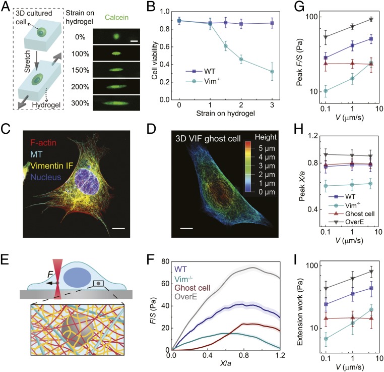
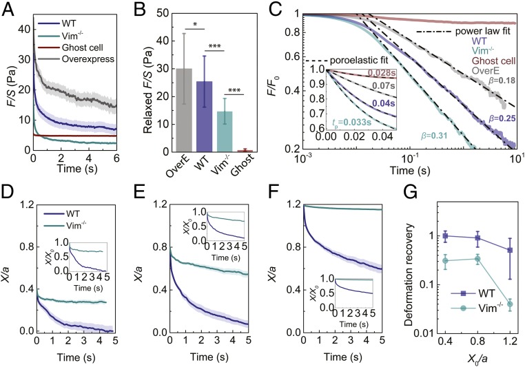
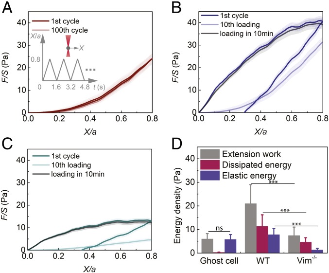
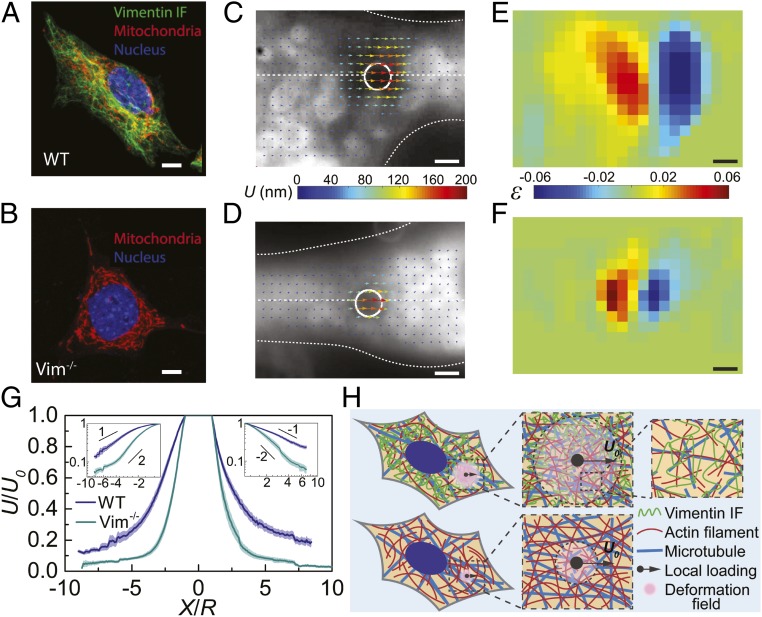
In many developmental and pathological processes, including cellular migration during normal development and invasion in cancer metastasis, cells are required to withstand severe deformations. The structural integrity of eukaryotic cells under small deformations has been known to depend on the cytoskeleton including actin filaments (F-actin), microtubules (MT), and intermediate filaments (IFs). However, it remains unclear how cells resist severe deformations since both F-actin and microtubules yield or disassemble under moderate strains. Using vimentin containing IFs (VIFs) as a model for studying the large family of IF proteins, we demonstrate that they dominate cytoplasmic mechanics and maintain cell viability at large deformations. Our results show that cytoskeletal VIFs form a stretchable, hyperelastic network in living cells. This network works synergistically with other cytoplasmic components, substantially enhancing the strength, stretchability, resilience, and toughness of cells. Moreover, we find the hyperelastic VIF network, together with other quickly recoverable cytoskeletal components, forms a mechanically robust structure which can mechanically recover after damage.

Keywords: cell mechanics; cytoplasm; cytoskeleton; intermediate filament; vimentin.


Conflict of interest statement

The authors declare no conflict of interest.

Figures

Fig. 1.
Fig. 1.
VIF network maintains cell viability under large deformations and increases the mechanical strength, stretchability, and toughness of the cytoplasm. (A) Cells cultured in 3D PEG-alginate hydrogels are deformed by stretching the hydrogels. Cells are labeled with calcein to visualize cell shape and viability. (Scale bar: 20 µm.) (B) The viability of WT and Vim−/− mEFs are measured with live/dead assay under various strains for 2 h (n = 15). (C) Immunofluorescence image of WT mEF showing the microtubule and vimentin IF cytoskeletons. (Scale bar: 10 µm.) (D) A 3D microscopic image of VIF in a ghost cell. The color represents height. (Scale bar: 10 µm.) (E) Schematic of micromechanical measurements in the cytoplasm using optical tweezers. The cytoskeletal mesh size and bead size are not in their actual proportion, just for illustration. (F) Normalized force–displacement curves obtained in cells at V = 1 µm/s, from which we calculate peak F/S, peak X/a, and extension work. The semitransparent band around the average curves represents the SE (n = 20 cells for each curve). (GI) The dependence of peak F/S (G), peak X/a (H), and extension work (I) on loading rate in different cells. Error bars represent SD (n = 20 cells). These mechanical properties have statistical difference between WT and Vim−/− mEFs and between WT and OverE mEFs (P < 0.001 in Student’s t test).
Fig. 2.
Fig. 2.
The elastic VIF network increases the relaxed force, relaxation time, and yielding strain of the cytoplasm. (A) Force relaxation curves of different cells under an instant initial X/a = 0.4. The semitransparent band around the average curves represents the SE (n = 15 cells for WT and overexpress, n = 25 cells for Vim−/− and ghost cell). (B) Comparison of cellular relaxed force, which is defined as the decrease of F/S over the relaxation test. Error bars represent SD (n = 15 cells for WT and overexpress, n = 25 cells for Vim−/− and ghost cell). (C) Relaxation curves are normalized with initial F0 at t = 0. The curves are fitted with viscoelastic power law decay at long time scales (0.05 s < t < 10 s) and are fitted with poroelastic exponential decay (Inset) at short time scales (t < 0.05 s). (DF) Deformation recovery in the cytoplasm of WT and Vim−/− mEFs under different initial normalized displacement (X0/a = 0.4, X0/a = 0.8, and X0/a = 1.2, respectively). The semitransparent band around the average curves represents the SE (n = 15 cells for each curve). (G) The cytoplasmic deformation recovery (defined as the recovered deformation over initial deformation) under different initial deformations in WT and Vim−/− mEFs. *P < 0.05; ***P < 0.001.
Fig. 3.
Fig. 3.
The VIF network increases the toughness of the cytoplasm by increasing dissipated energy and elastic energy. (A) Cyclic loading in the cell is achieved by reciprocating movement of a bead at a speed of 1 µm/s using optical tweezers (Inset). The first and 100th loading and unloading curves in VIF ghost cells overlap. The semitransparent band around the average curves represents the SE (n = 15 cells). (B and C) Plot of the first and 10th cyclic loading curves in WT mEFs (B) and Vim−/− mEFs (C). After being damaged by repeated loadings, the loading curves in both WT and Vim−/− mEFs recover to original levels in 10 min, highlighting the self-healing nature of the cytoplasm. The semitransparent band around the average curves represents the SE (n = 20 cells for each curve). See full curves in SI Appendix, Fig. S10. (D) VIF ghost cell does not dissipate energy. WT mEFs have significantly higher extension work, dissipated energy, and elastic energy than Vim−/− mEFs. Error bars represent SD (n = 15 cells for ghost cells, n = 20 cells for WT and Vim−/− mEFs). ns, not significant; ***P < 0.001.
Fig. 4.
Fig. 4.
VIFs extend the cytoplasmic deformation field under local loading, acting as a stretchable hyperelastic network. (A and B) Deformation field is obtained by dragging a 2-µm-diameter bead in the cytoplasm over 200 nm and visualizing the 2D projected movements of surrounding fluorescently labeled mitochondria. (C and D) The displacement fields around the loading beads (white circle) in WT and Vim−/− mEFs (cell boundaries are marked with white dotted lines). The color and the length of arrows represent displacement magnitudes. (E and F) The normal strain fields in WT and Vim−/− mEFs are obtained from displacement fields shown in C and D. The color represents the magnitude of normal strain. (G) Plot of the normalized cytoplasmic displacement as a function of the normalized distance to the loading center, along the drag directions (horizontal white dashed lines in C and D). Log-log plots are shown in the Insets. The semitransparent band around the average curves represents the SE (n = 15 cells for each curve). (H) Schematics to illustrate the mechanism of extending deformation fields by VIF network. Highly stretchable VIF networks maintain the elasticity of and transmit local strain through a large zone in the cytoplasm, while other cytoskeletal structures are easy to be damaged under deformation. The cytoskeletal mesh size and bead size are not in their actual proportion, just for illustration. (Scale bars: 10 µm in A and B and 2 µm in CF.)

References

    1. Kalluri R., EMT: When epithelial cells decide to become mesenchymal-like cells. J. Clin. Invest. 119, 1417–1419 (2009). - PMC - PubMed
    1. Yang J., Weinberg R. A., Epithelial-mesenchymal transition: At the crossroads of development and tumor metastasis. Dev. Cell 14, 818–829 (2008). - PubMed
    1. Mantovani A., Cancer: Inflaming metastasis. Nature 457, 36–37 (2009). - PubMed
    1. Denais C. M., et al. , Nuclear envelope rupture and repair during cancer cell migration. Science 352, 353–358 (2016). - PMC - PubMed
    1. Raab M., et al. , ESCRT III repairs nuclear envelope ruptures during cell migration to limit DNA damage and cell death. Science 352, 359–362 (2016). - PubMed

Publication types

LinkOut - more resources