. 2019 Oct;38(44):6959-6969.
doi: 10.1038/s41388-019-0913-4. Epub 2019 Aug 13.

Betaglycan drives the mesenchymal stromal cell osteogenic program and prostate cancer-induced osteogenesis

Affiliations

Betaglycan drives the mesenchymal stromal cell osteogenic program and prostate cancer-induced osteogenesis

Leah M Cook et al. Oncogene. 2019 Oct.

Abstract

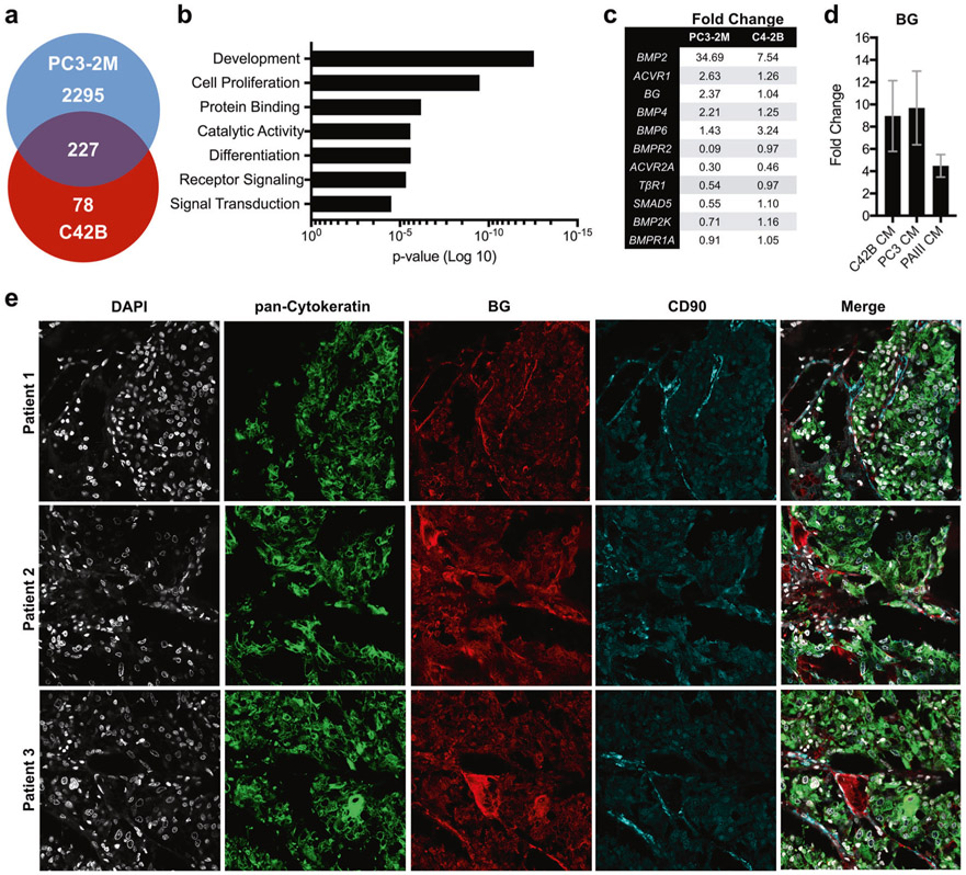
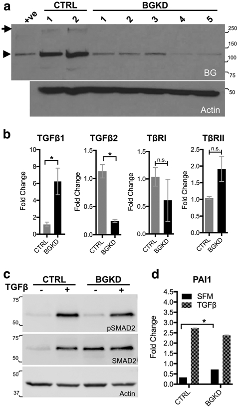
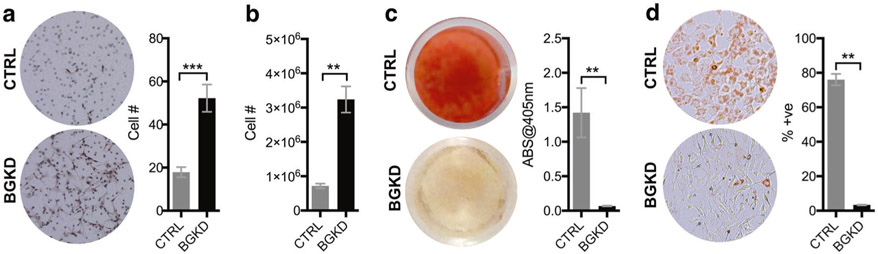
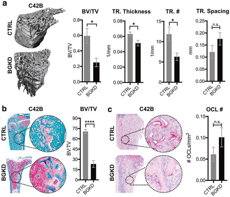
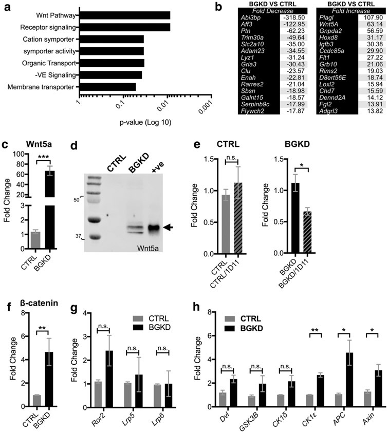
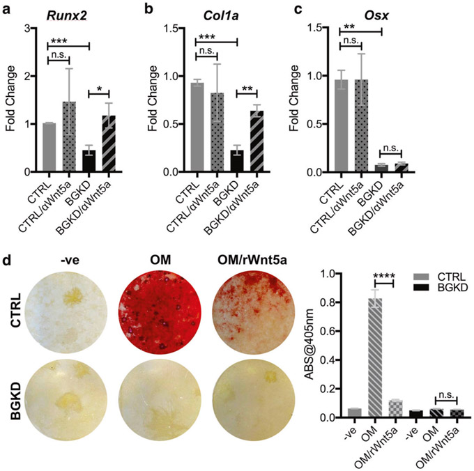
Bone metastatic prostate cancer provokes extensive osteogenesis by driving the recruitment and osteoblastic differentiation of mesenchymal stromal cells (MSCs). The resulting lesions greatly contribute to patient morbidity and mortality, underscoring the need for defining how prostate metastases subvert the MSC-osteoblast differentiation program. To gain insights into this process we profiled the effects of co-culture of primary MSCs with validated bone metastatic prostate cancer cell line models. These analyses revealed a cast of shared differentially induced genes in MSC, including betaglycan, a co-receptor for TGFβ. Betaglycan has not been studied in the context of bone metastatic disease previously. Here we report that loss of betaglycan in MSC is sufficient to augment TGFβ signaling, proliferation and migration, and completely blocks the MSC-osteoblast differentiation program. Further, betaglycan was revealed as necessary for prostate cancer-induced osteogenesis in vivo. Mechanistically, gene expression analysis revealed betaglycan controls the expression of a large repertoire of genes in MSCs, and that betaglycan loss provokes >60-fold increase in the expression of Wnt5a that plays important roles in stemness. In accord with the increased Wnt5a levels, there was a marked induction of canonical Wnt signaling in betaglycan ablated MSCs, and the addition of recombinant Wnt5a to MSCs was sufficient to impair osteogenic differentiation. Finally, the addition of Wnt5a neutralizing antibody was sufficient to induce the expression of osteogenic genes in betaglycan-ablated MSCs. Collectively, these findings suggest a betaglycan-Wnt5a circuit represents an attractive vulnerability to ameliorate prostate cancer-induced osteogenesis.


Conflict of interest statement

Conflict of interest The authors declare that they have no conflict of interest.

Figures

Fig. 1
Fig. 1
Prostate cancer cells induce betaglycan expression in bone marrow-derived MSCs. a Microarray analysis of differential gene expression induced in bone marrow MSCs in response to culture in C42B-conditioned and PC3-conditioned media (CM, 6h). b Gene ontology analysis of biological processes. c Expression of TGFβ pathway members in MSCs that are regulated by culture in CM from PC3-2M and C4-2B cells. d qRT-PCR analysis of betaglycan (BG) mRNA levels in MSCs after 6 h incubation in CM derived from C4-2B and PC3-2M, and from CM from the independent rodent prostate cancer cell line, PAIII. Data shows fold increase over basal BG expression. e Betaglycan (BG; red) localization in human bone metastatic prostate cancer specimens (n = 10). Representative images from three individual patient specimens are illustrated. CD90 (blue) was used to localize MSCs while pan-cytokeratin (green) was used to localize the prostate cancer metastases. DAPI (gray) was used as a nuclear counterstain
Fig. 2
Fig. 2
Betaglycan knockdown enhances TGFβ signaling in MSCs. a Analysis of Betaglycan protein levels in scrambled control (CTRL) and betaglycan CRISPR ablated (BGKD) clonal MSC populations. Arrow indicates full-length betaglycan protein while arrowhead indicates the core protein. Recombinant BG was used as a positive (+ve) control while the actin immunoblot was used as a loading control. b qRT-PCR analysis of TGFβ ligand isoforms (TGFβ1 and TGFβ2) and TGFβ receptor I and II (TβRI, TβRII) mRNA levels in betaglycan knockdown (BGKD) MSCs normalized to those in control (CTRL) MSCs. c Phosphorylated SMAD2 (pSMAD2) at basal levels (−) and in response to TGFβ treatment (+, 5 ng/ml for 30 min). Actin was used as a loading control. d PAI1 promoter-reporter outputs at basal levels in serum-free media (SFM) and in response to TGFβ treatment (5 ng/ml for 24 h). Molecular weights are in kDa
Fig. 3
Fig. 3
Betaglycan inhibits MSC migration and proliferation and is necessary for osteogenic and adipogenic MSC differentiation. a The number of migrating CTRL and BGKD MSCs in serum-free media was measured over a 5 h period using a Boyden chamber. Representative images from each group are shown. b CTRL and BGKD cell proliferation at 7 days was measured using cell counts. c Representative images of Alizarin Red staining for mineralization in CTRL and BGKD MSCs incubated with osteogenic media for 21 days. The amount of solubilized Alizarin Red was also measured in CTRL and BGKD MSCs. d The number of Oil Red O-positive cells as a function of total number of cells was measured in CTRL and BGKD MSCs incubated for 3 days in adipogenic media. Representative images from each group are shown. Asterisks denote statistical significance (**p < 0.01)
Fig. 4
Fig. 4
Betaglycan controls prostate cancer-induced osteogenesis. a Representative μCT images of tibias 7 weeks following co-inoculation with C4-2B prostate cancer cells and CTRL or BGKD MSCs. Graphs illustrate μCT values in each group for bone volume as function of total volume (BV/TV), trabecular thickness (TR. Thickness), trabecular number (TR. #), and trabecular spacing (TR. Spacing). b, c Representative images of trichrome b and TRAP c stained sections derived from CTRL/C4-2B and BGKD/C4-2B groups. BV/TV ratios b and osteoclast numbers/mm of tumor-bone interface (OCL#; red) were quantitated in non-sequential sections derived from each group. Asterisks denote statistical significance (*p < 0.05, ****p < 0.001) while n.s. indicates non-significance
Fig. 5
Fig. 5
Betaglycan controls Wnt5a expression in MSCs. a Gene ontology analysis of microarray data obtained from CTRL and BGKD MSC grown under normal serum-containing culture conditions. b Microarray analysis of average gene expression identified the top-15 up-regulated and down-regulated genes in the BGKD MSCs compared to CTRL. c, d qRT-PCR c and immunoblot d analysis of Wnt5a expression in CTRL and BGKD MSCs. Arrow indicates Wnt5a. Recombinant Wnt5a was used as a positive control (+ve). Molecular weights shown are in kDa. e qPCR of Wnt5a mRNA levels following incubation of CTRL and BGKD MSCs with the TGFβ inhibitor 1D11 (10 μg/ml for 6 h). 1D11 effects are normalized relative to respective non-treatment controls. fh qPCR analysis of β-catenin f, Wnt receptors g and Wnt signaling effectors h at baseline in CTRL and BGKD MSCs. Asterisks denote statistical significance (*p < 0.05, **p < 0.01, ***p < 0.005) while n.s. denotes non-significance
Fig. 6
Fig. 6
Betaglycan regulation of Wnt5a contributes to the MSC-osteoblast differentiation program. a–c qPCR analysis of RUNX2 (RUNX2) a, α-type I collagen (COL1A1; b), and osterix (OSX; c) in CTRL and BGKD MSCs treated in the presence or absence of a Wnt5a blocking antibody (αWnt5a; 1 μg/ml; 6 h). Fold changes are normalized to each respective non-treated control. d Representative images of Alizarin Red staining for mineralization in CTRL and BGKD MSCs incubated with osteogenic media (OM) for 21 days in the presence or absence of recombinant Wnt5a (rWnt5a; 0.5 μg/ml). Cells were also treated with non-OM media as a negative control (−ve). The amount of solubilized Alizarin Red was measured in the treated CTRL and BGKD MSCs. Asterisks denote statistical significance (**p < 0.01, ****p < 0.0001)

Comment in

References

    1. Logothetis C, Morris MJ, Den R, Coleman RE. Current perspectives on bone metastases in castrate-resistant prostate cancer. Cancer Metastas-Rev. 2018;37:189–96. - PMC - PubMed
    1. Frieling JS, Basanta D, Lynch CC. Current and emerging therapies for bone metastatic castration-resistant prostate cancer. Cancer Control: J Moffitt Cancer Cent. 2015;22:109–20. - PMC - PubMed
    1. Cook LM, Shay G, Aruajo A, Lynch CC. Integrating new discoveries into the “vicious cycle” paradigm of prostate to bone metastases. Cancer Metastas-Rev. 2014;33:511–25. - PMC - PubMed
    1. Ren G, Esposito M, Kang Y. Bone metastasis and the metastatic niche. J Mol Med. 2015;93:1203–12. - PMC - PubMed
    1. Juarez P, Guise TA. TGF-beta in cancer and bone: implications for treatment of bone metastases. Bone. 2010;48:23–29. - PubMed

Publication types

MeSH terms